Body planes and sections
A plane is an imaginary flat surface passed through the body, and a section is what that plane exposes. This chapter names the three standard planes, shows why the same plane meets different organs at different levels, and teaches you to read the direction letters printed on any image.
By the end of this chapter you can
- Name the three standard planes, give the synonyms transverse and frontal, and state which body portions each plane separates.
- Tell a midsagittal plane from a parasagittal plane and name organs that only one of them can cross.
- Explain why two sections in the same plane can show different organs, and name what an axial plane meets at the nipples, the navel, and the hips.
- Explain what a single section can and cannot show, and why neighboring sections are needed to follow a tube-shaped structure such as the aorta.
- Read the letters R, L, A, P, S, and I on an image as the patient's own directions, recognize an oblique plane, and tell a long-axis view of an organ from a short-axis view.
Three planes, three ways to cut
A is an imaginary flat surface passed through the body, like a pane of glass. Anatomy names three standard planes that each cut through the body in a different direction.
An plane, also called a plane, cuts straight across the long axis of the body. When a person is standing in anatomical position, the axial plane lies parallel to the floor. The axial plane divides the body into superior and inferior portions, like a cut across a loaf of bread.
A plane, also called a plane, stands upright and runs from side to side, passing from one ear to the other like a crown. It divides the body into anterior and posterior portions.
A plane also stands upright, but it runs from front to back, like an arrow passing through the nose and out the back of the head. It divides the body into right and left portions. Sagitta is the Latin word for arrow.
Each plane separates exactly one pair of directions. An axial plane separates superior (above) from inferior (below). A coronal plane separates anterior (front) from posterior (back), and a sagittal plane separates right from left.
Like directions, planes travel with the body. When a patient rolls from to , the coronal plane through the chest still separates anterior from posterior.
In the figure below, choose Axial, Coronal, or Sagittal to see that plane on its own.
Interactive model
Three planes through one body
All three planes meet in the chest. Pick one to see the section it makes, then move it.
Three planes at right angles. Each one separates one pair of directions. The dashed lines show where the other two planes cross each section.

Body location map
Midsagittal and parasagittal
The plane, also known as the median plane, is the one sagittal plane that runs from anterior to posterior directly through the of the body. It divides the body into equal right and left halves.
Every other sagittal plane runs parallel to the midsagittal plane but lies to either side of it. These are known as planes, where para means beside. A plane only two centimeters from the midline is parasagittal, and so is a plane fifteen centimeters from it. Any sagittal plane that does not pass directly through the midline is parasagittal.
The midsagittal plane passes through structures that sit directly on the midline, such as the sternum (the breastbone), the vertebral column (the spine), and the urinary bladder. The midsagittal plane misses every paired organ, like the eyes, lungs, and kidneys. These paired organs sit lateral to the midline, one on each side.
Slide the plane six centimeters toward the patient's right and it becomes parasagittal. It now passes through the right lung, the liver, and the right kidney. Since the plane is no longer midsagittal, it passes lateral to the spine.
In many areas the two sides of the body are near mirror images of each other. A parasagittal section through the right side can look much like one through the left. The letters printed on the image always refer to the patient, which makes them the surest way to tell the two sides apart.
Same plane, different level
Every slice of a loaf of bread faces the same way, yet each slice shows a different part of the loaf. The same is true of body planes: the type of plane gives the direction, and the gives the plane's position along the body. A level is named by a the plane passes through. For example, an axial plane at the level of the kidneys or a coronal plane through the hip joints gives the precise location of an image.
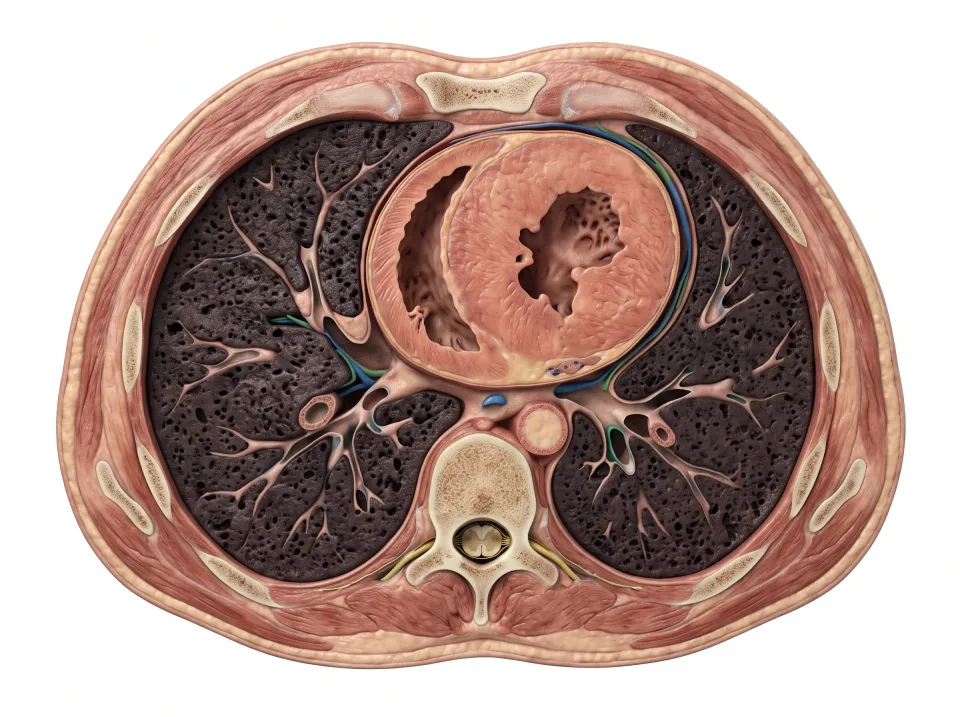
At the level of the nipples, an axial plane passes through the chest and cuts the heart with a lung lateral to it on each side. Move that axial plane inferiorly to the upper abdomen, and it meets the liver on the patient's right and the stomach on the patient's left.
Continue moving the plane inferiorly to the level of the navel (the belly button), and it meets the kidneys. It also crosses through the and , the body's largest artery and vein, which lie anterior to the spine. At the level of the hips, inside the pelvis, it meets the urinary bladder anteriorly and the rectum (the last part of the bowel) posteriorly.
Parallel axial planes are stacked from superior to inferior, parallel coronal planes from anterior to posterior, and parallel sagittal planes side by side from right to left. Move the plane through the transparent body below and watch which organs light up at each level.
Interactive model
Move the plane through the organs
Slide the plane or jump to a landmark and watch the section change. Then try the three tasks.
lies flat, like slicing a loaf of bread. It separates upper from lower.
Body location map
What this slice shows
Seen from the feet, the way axial images are shown, so the patient's right is on your left.
Axial through the heart
Try it
0 of 3 done
What one section can and cannot show
A is the flat surface a plane exposes at one position. It shows only the structures the plane touches at that point. A structure that is missing from one axial section may simply lie superior or inferior to it.
Because a section is flat, it can mislead you about a structure's shape. A sausage sliced across shows a small circle, and the same sausage sliced lengthwise shows a long strip. The aorta is a long tube that runs from superior to inferior down the trunk. On an axial section it is a small circle anterior to the spine, and on a coronal or sagittal section it is a long stripe running along the trunk. A circle in one section can therefore be a long tube passing through the plane.
For this reason, never judge a structure from a single . Follow it through the neighboring sections and watch how the structure changes. A tube such as the aorta stays a circle from one section to the next. On axial sections the kidney is a small oval near its superior end, widens through its middle, and narrows again toward its inferior end. Scrolling through the sections in this way rebuilds the three-dimensional organ in your mind.
Sagittal: the aorta as one long stripe
Axial: five neighbors, top to bottom
A ring in front of the spine at every level. Same size, same place: a tube passing through each plane.
Reading the letters on an image
Images carry small letters at their edges, and each letter names a direction on the patient. R and L are the patient's right and left, A and P are anterior and posterior, and S and I are superior and inferior. Some systems print H and F, for head and feet, in place of S and I.
An axial image is displayed as if you were standing at the patient's feet and looking toward the head. The patient's right therefore appears on the left side of the picture, and the R marker sits at the left edge. This is why the liver, which lies on the patient's right, appears on the left of an axial image.
The letters also tell you which plane you are looking at. An axial image has R and L on its sides, with A at the top and P at the bottom. A coronal image has R and L on its sides, with S at the top and I at the bottom. It is displayed as if you were facing the patient, so the patient's right appears on the left of the picture. A sagittal image has A and P on its sides, with S at the top and I at the bottom. Read the letters before you name any structure, because an image can be flipped or turned while the letters stay attached to the patient.

Oblique planes
An plane is a plane tilted away from all three standard planes. Oblique planes are useful because many organs sit at an angle inside the body, and the standard planes cut them crookedly.
The heart lies tilted, with its tip pointing inferiorly and to the patient's left. Each kidney leans so that its superior end is more medial than its inferior end. The spine curves, and the discs between its bones, the vertebrae, follow that curve. A plane angled to match one of these structures shows it in one clean piece, while the standard planes scatter it across several sections.
Oblique planes are described with the same words as the standard planes. Each one is tilted from axial, coronal, or sagittal, and its level is named by a landmark it passes through.
Sliding a plane and tilting it are different moves. Sliding keeps the plane's direction and changes only its level, so an axial plane stays axial. Tilting changes the direction itself, and that change is what makes a plane oblique.
Long axis and short axis
An organ that sits at an angle has its own axis. This axis is a line that runs along the organ's length. A plane lined up with that axis gives a long-axis view, and a plane at a right angle to the axis gives a short-axis view. A banana shows the difference: a lengthwise cut gives one long strip, and a cut across gives a circle. Both are called oblique planes, named for the organ's own axis.
The heart lies tilted in the chest, with its tip pointing inferiorly, anteriorly, and to the patient's left. The standard planes would cut the heart at awkward angles. A long-axis view follows the heart from its base to its tip and shows its chambers end to end. The heart's two main pumping chambers are the left and right . A short-axis view cuts straight across both ventricles: the thick wall of the left ventricle shows as a ring, with the thinner right ventricle curved against it like a crescent.
Optional: a look ahead to MRI
A first look at MRI
Four MRI examples to browse: each shows its plane and the anatomy it crosses.

sagittal plane
Brain (midline)
The brain in profile. The face points to the viewer's left, the cerebellum tucks under the back of the brain, and the brainstem runs down toward the spinal cord. Anterior to posterior and superior to inferior are both in the picture, so this is sagittal.
Midline sagittal slice; anterior (face) is to the left.
Practice
Read a section
Six quick questions on sections cut from the model: tap an organ, name a plane from its letters, or name the level. Practice only, nothing is graded.
Key ideas to remember
- (transverse) separates superior from inferior, (frontal) separates anterior from posterior, and separates right from left.
- Only the plane splits the body into equal halves. Every other sagittal plane is and can cross paired organs such as a lung or a kidney.
- The plane gives the direction and the level gives the location, so an axial plane at the nipples meets the heart while the same plane at the hips meets the bladder.
- A section shows only what the plane touches, so follow a structure through neighboring sections. Read the R, L, A, P, S, and I letters as the patient's own directions.
Check what you learned
Twenty-five questions from this chapter, two of them illustrated, each with the answer, the reasoning, and a memory aid.
1 of 4: The patient lies supine. Seen from above with the head at the top. The patient's right hand is on your left, and so is the liver.
1 of 5: Axial. The plane lies flat, parallel to the floor.