Bones, joints, and muscles
Bones support and protect the body, joints connect two or more bones together, and muscles produce the movement of the body. Learn the major bones and the relationships that make everyday movement possible.
By the end of this chapter you can
- Sort the major bones into the axial and appendicular skeletons, and name the bones of each limb from proximal to distal.
- State the vertebral counts for each region, tell the atlas from the axis, and name a disc level such as L4-L5 correctly.
- Describe the three joint types and the parts of a synovial joint, including the menisci and the four main ligaments of the knee.
- Use flexion, extension, abduction, adduction, rotation, supination, pronation, dorsiflexion, and plantar flexion to describe how a patient's limb is positioned.
- Tell a tendon from a ligament, explain why muscles work in opposing pairs, and name the muscle groups that matter when you position a shoulder, knee, or ankle.
Bone is living tissue
Bone is living that is made of cells and blood vessels set in a hard matrix that is stiffened with calcium and other minerals. The main jobs of the skeletal system include keeping the body upright, protecting the brain, heart, and lungs, and storing calcium that can be moved in and out of the bloodstream. The marrow located inside bone also makes the body's blood cells.
is the dense, hard outer shell of a bone. Inside it, forms a sponge of thin struts that keeps the bone light but strong. The spaces between the struts hold the marrow.
makes red blood cells, white blood cells, and platelets. In a child, red marrow is found in most bones. In an adult, red marrow is concentrated in the spine, pelvis, ribs, sternum, and the ends of the femur and humerus. The marrow found in the rest of the adult skeleton has turned to fatty yellow marrow.
Marrow is why bone is such a rewarding tissue on MRI. The scanner sees the fat and water inside a bone far better than it sees cortical bone, which appears as a thin dark line. As a result, a bruise, an infection, or a tumor inside a bone can show on MRI while an X-ray still looks normal.
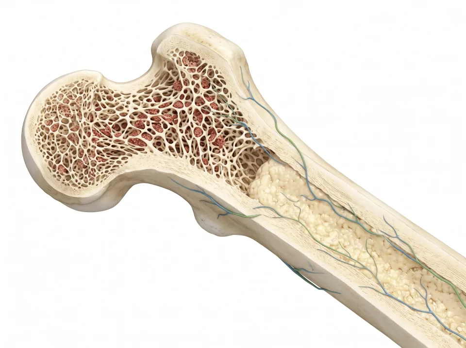
1.Cortical shell
A dense outer shell provides strength and protection.
2.Trabecular lattice
An inner network supports loads while leaving spaces for marrow.
3.Marrow
Red marrow makes blood cells. Yellow marrow contains more fat.
Two skeletons in one: axial and appendicular
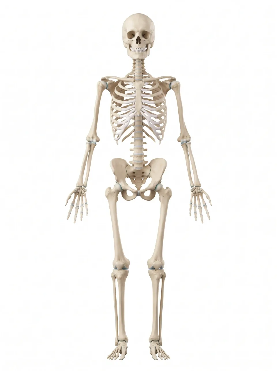
Anatomists divide the roughly 206 adult bones into two groups. The is located along the body's central axis. The axial skeleton consists of the skull, the , and the thoracic cage (the sternum and twelve pairs of ribs).
The is everything that hangs off the axial skeleton. It consists of the upper and lower limbs, along with the shoulder and pelvic girdles that attach them to the trunk. The shoulder girdle is made of two bones, the (shoulder blade) and the (collarbone). The pelvic girdle is made of the two hip bones, and each hip bone is three bones fused together: the ilium, ischium, and pubis.
The skull sits at the superior end of the axial skeleton. It consists of the cranium (the bony protective barrier around the brain) and the bones of the face. The mandible (lower jaw) is the one skull bone that moves freely. It hinges at the temporomandibular joint, just anterior to the ear. If you rest a finger there and open your mouth, you can feel the jaw move.
The vertebral column runs inferiorly from the skull to the pelvis and supports the head and trunk. The (breastbone) lies in the anterior midline of the chest and anchors the ribs. The ribs curve around the chest to meet the posteriorly, and together these bones form the cage that protects the heart and lungs.
Interactive model
The whole skeleton is highlighted. Turn it to trace the axial skeleton down the center (skull, vertebral column, ribs, and sternum), then the appendicular skeleton: the two girdles and the limbs.
Body location map
Highlighted structure
Skeleton
Where: Central framework and limbs
Job: Supports movement, protects organs, and stores minerals.
Drag to turn. Pinch to zoom. Select a visible structure to learn about it.
Where it sits on a slice
Coronal through the skeleton
Seen from the front, so the patient's right is on your left.
The bones of the limbs
The joins the superior end of the sternum to the at the point of the shoulder, and the scapula lies flat against the posterior rib cage.
The , the single bone of the upper arm, meets the scapula at the shoulder. The forearm has two bones: the on the thumb side and the on the side of the little finger. The proximal radius and ulna meet the humerus to form the elbow joint, and distally at the wrist the radius meets the eight small bones known as the carpals. Distal to the carpals lie the five metacarpals of the palm and the phalanges of the fingers.
In each hip bone, the ilium is the broad superior wing, the ischium the inferior part you sit on, and the pubis is the anterior part. The three fuse during adolescence, and the hip bones meet the posteriorly to form the pelvis.
The is the longest bone in the body. It meets the hip bone proximally to form the hip joint and the distally to form the knee joint. The (kneecap) lies anterior to the distal femur, inside the of the thigh muscles. Distal to the knee, the thick tibia (shinbone) carries the weight and lies to the slender on the side of the leg. At the ankle, both bones meet the talus, one of the seven tarsals. Another tarsal, the calcaneus, forms the heel, and distal to the tarsals lie the five metatarsals and the phalanges of the toes.
1.Axial skeleton
Skull, spine, ribs, and sternum form the central framework.
2.Upper limb
Clavicle and scapula form the shoulder girdle; humerus is the upper arm bone.
3.Forearm
In anatomical position, radius is on the thumb side; ulna is on the little-finger side.
4.Lower limb
Femur is in the thigh. Tibia is the larger medial shin bone; fibula is the slender lateral bone.
The vertebral column, level by level
The is a stack of 33 in five regions, and every spine MRI scan is ordered by region. Seven vertebrae (C1 to C7) form the neck, and twelve vertebrae (T1 to T12) each carry a pair of ribs. Five vertebrae (L1 to L5) are the largest, because they carry the most weight, and they form the small of the back. Inferior to them, five vertebrae are fused into the , and three to five tiny ones form the .
C1 is known as the atlas. It is a ring-shaped vertebra that holds up the skull, as Atlas of the myth held up the sky. C2 is called the axis and has a peg that C1 pivots around when you shake your head.
Each bump you can feel along the midline of the back is the spinous process of one vertebra. The big bump at the base of the neck is C7.
An sits between each pair of vertebral bodies, from C2-C3 to L5-S1. Each disc is a tough fibrous ring around a soft gel center, and it spreads load and allows the spine to bend. A disc takes the name of the two vertebrae it sits between, so the disc between the fourth and fifth lumbar vertebrae is L4-L5. A vertebra gets a single label, such as L4.
The spinal cord runs inside the canal that the vertebrae form, but it ends near L1-L2. The cord and the inferior to it belong to the nervous system chapter.
On a sagittal image of the spine, the vertebral bodies stack like blocks with a disc between each pair. You name a level by counting from a known end: inferiorly from C2 at the superior end of the neck, or superiorly from the sacrum. The iliac crests, the superior edges of the hip bones where you rest your hands, are a landmark you can feel: a line joining their most superior points crosses the spine at about the fourth lumbar vertebra.
Cutting the section…
Tap a vertebral body, or the disc between two of them, to name it. Or step through the levels with the arrows.
Try it
- Find C7, the bump at the base of the neck. Not done yet.
- Find T12, the last vertebra that carries a pair of ribs. Not done yet.
- Find the L4-L5 disc. Not done yet.
Interactive model
The vertebral column is highlighted from the back. Choose Left or Right to follow it from the base of the skull down to the sacrum.
Body location map
Highlighted structure
Vertebral column
Where: Along the back, from the neck to the pelvis
Job: Supports the trunk and protects the spinal cord and nerve roots.
Drag to turn. Pinch to zoom. Select a visible structure to learn about it.
Where it sits on a slice
Sagittal through the vertebral column
Seen from the patient's left, with the face toward your left.
Where bones meet: three kinds of joint
A joint (articulation) is any place two bones meet, and how much a joint moves depends on what lies between them. are held by tough connective tissue and barely move. The sutures between the flat bones of the skull are the classic example. They are nearly rigid seams in an adult.
have a pad of cartilage between the bones and allow a little movement. Examples are the intervertebral discs and the , where the two pubic bones meet anteriorly.
are the freely movable ones: shoulder, elbow, wrist, hip, knee, ankle, fingers, and toes. Each bone end is capped with , which is a smooth, glassy layer that lets the surfaces glide with little friction. A fibrous capsule wraps the joint, and its inner lining, the synovial membrane, secretes synovial fluid, which lubricates and feeds the cartilage. are thick bands that connect bones, reinforce the capsule, and keep the joint from moving in directions it should not.
The knee adds two , C-shaped pads of fibrocartilage between the femur and tibia that spread load and deepen the fit. It also has four main ligaments. The anterior and posterior cruciate ligaments cross in the center of the joint, and the medial and lateral collateral ligaments brace its sides. Injuries to those menisci and four ligaments are the most common reasons knee MRIs are ordered.
The shoulder and hip have a , a rim of cartilage that deepens the socket. Small fluid-filled sacs called sit where tendons rub over bone to reduce friction.
On a sagittal image of the knee, each meniscus shows as a dark triangle wedged between the femur and the tibia. On the slices nearest the medial or lateral edge of the knee, its anterior and posterior parts join into a bow-tie shape. The cruciate ligaments show as dark bands crossing in the notch at the center of the knee.
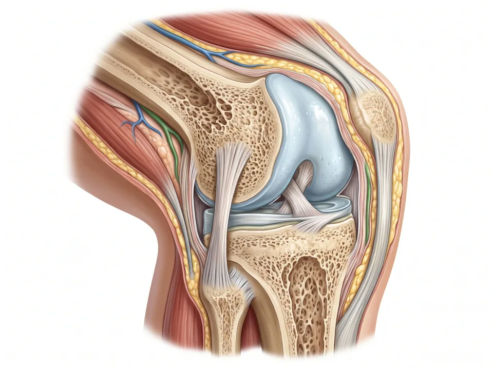
1.Cartilage
Smooth cartilage covers the bone ends and helps them glide.
2.Joint space
A small space containing synovial fluid reduces friction; a capsule encloses the joint.
3.Ligament
A ligament joins bone to bone and helps stabilize the joint.
A knee is one example of a synovial joint. The joint space is small and contains fluid.
The language of movement
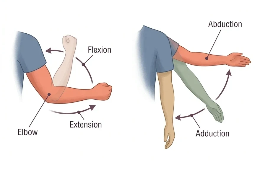
Movement words describe what happens at a joint. You use them to tell a patient how to hold a limb and a colleague how it was positioned. They come in pairs, and each word has a partner that names the opposite movement.
turns a bone around its own long axis, as the humerus does when the whole arm turns at the shoulder. and turn only the forearm and happen where the radius rolls over the ulna, not at the shoulder.
Try each movement in the table on yourself and say its name aloud.
| Term | What happens | Example |
|---|---|---|
| Decreases the angle at a joint | Bending the elbow | |
| Increases the angle at a joint | Straightening the elbow again | |
| Moves a limb away from the midline | Lifting the arm out to the side | |
| Brings a limb back toward the midline | Lowering the arm back to the side | |
| Lateral (external) | Turns the front of the limb outward | Turning the whole arm so the thumb points outward |
| Medial (internal) | Turns the front of the limb inward | Turning the whole arm so the thumb points inward |
| Turns the palm to face forward | Turning a bent forearm so the palm faces up | |
| Turns the palm to face backward | Turning a bent forearm so the palm faces down | |
| Pulls the top of the foot toward the shin | Standing on the heels | |
| Points the foot and toes down | Standing on the balls of the feet like a dancer |
Three kinds of muscle
The chapter on how the body is organized introduced three types of muscle. is striated (striped), attaches to bone, and works under conscious control.
and both work without conscious control. Cardiac muscle is also striated, but it is found only in the wall of the heart, where it beats to its own rhythm. Smooth muscle has no stripes and lines hollow organs and blood vessels, where it moves food along the gut and narrows or widens arteries. The panel below shows the three types side by side.
Of the three types, this chapter focuses on skeletal muscle. The body has more than 600 skeletal muscles, and together they make up about 40 percent of body weight. As a result, muscle is the largest tissue on almost any slice through a limb. Each muscle is a bundle of long fibers wrapped in connective tissue that gathers at both ends into a . A muscle can pull, but it cannot push.
Skeletal muscle is also the muscle a patient can hold still on request. That is why a shoulder or knee that stays motionless gives a sharp image, while the heart and gut keep moving on their own.
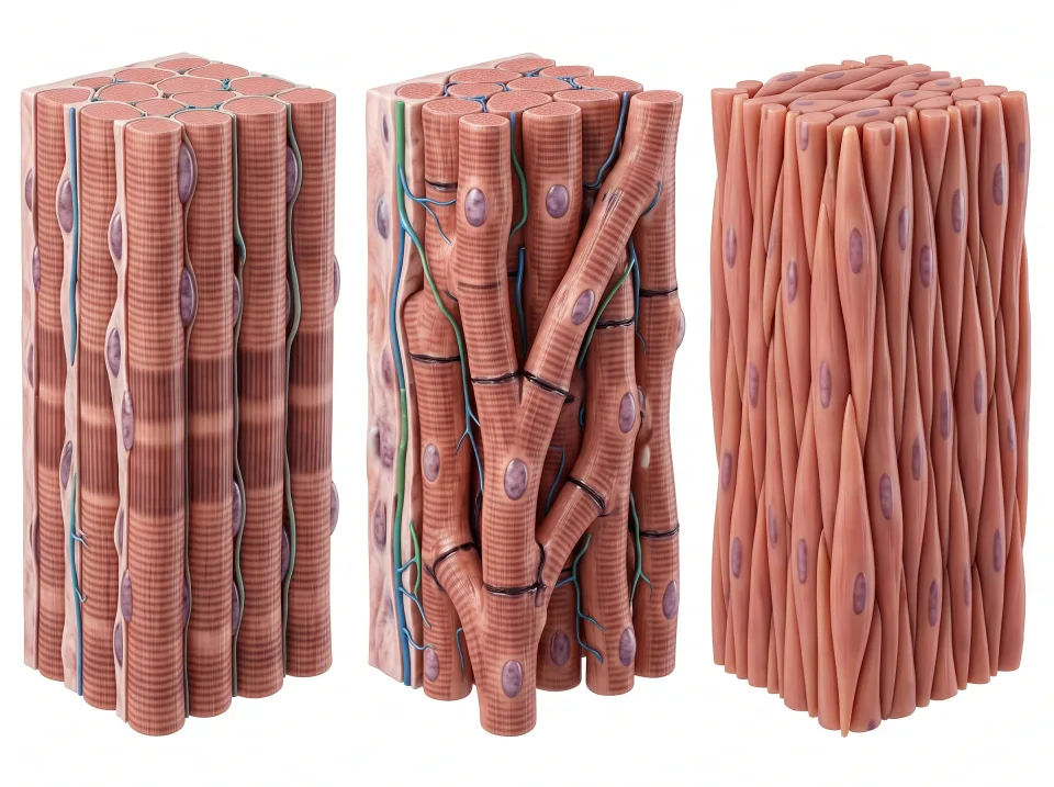
1.Skeletal
Attached to the skeleton: voluntary movement and posture.
2.Cardiac
In the heart wall: rhythmic pumping without conscious commands.
3.Smooth
In hollow organs and vessels: moving contents and changing tube diameter.
How a muscle moves a bone
When a skeletal muscle contracts, it shortens and develops tension. A carries that pull across a joint to the bone on the other side, so the bone swings toward the muscle. A is different: it runs from bone to bone, supports the joint, and pulls on nothing. Tendons tie muscle to bone, and ligaments tie bone to bone.
Because a muscle can only pull, joints are worked by opposing groups. One group pulls the joint one way, and its partner on the opposite side pulls it back. Together they can move the joint in either direction and hold it steady in any position.
At the shoulder, the caps the joint and lifts the arm out to the side (abduction). Deep to it, the four small muscles of the send their tendons around the head of the humerus and hold it in its shallow socket.
At the knee, the on the anterior thigh straighten (extend) the joint. They pull on the tibia through the quadriceps tendon, the patella, and the patellar ligament (which many clinicians still call the patellar tendon). The on the posterior thigh bend (flex) the knee.
At the ankle, the and the soleus deep to it form the calf. They point the foot down (plantar flexion) through the Achilles tendon, the thick cord you can pinch just superior to the heel. The tendon attaches to the posterior surface of the calcaneus.
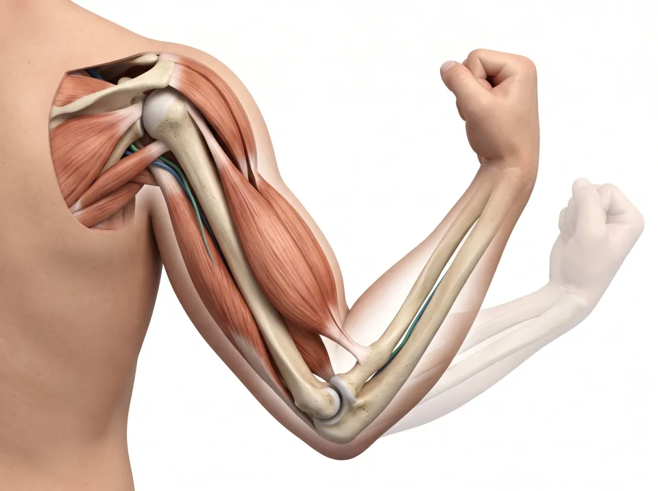
1.Muscle
Muscle develops tension when activated.
2.Tendon
A tendon transfers the muscle’s pull to bone.
3.Movement
The bone moves at a joint. An opposing muscle group helps return and control it.
Key ideas to remember
- The is the skull, spine, sternum, and ribs. The limbs and their girdles are .
- The spine has seven cervical, twelve thoracic, and five lumbar vertebrae, followed by the sacrum and coccyx. A disc carries both neighbors' names, such as L4-L5.
- Fibrous joints barely move, cartilaginous joints move a little, and synovial joints move freely on a cap of articular cartilage inside a fluid-filled capsule.
- Tendons tie muscle to bone, and ligaments tie bone to bone. A muscle can only pull, so muscles work in opposing pairs.
Check what you learned
Twenty-five questions from this chapter, two of them illustrated, each with the answer, the reasoning, and a memory aid.
1 of 4: Supination. Palm faces forward, the thumb points away from the body, and the radius and ulna lie side by side.
1 of 4: Relaxed. The arm hangs straight. The biceps in front of the upper arm and its partner behind are both at rest.