The nervous and endocrine systems
The nervous and endocrine systems coordinate the body's functions. Follow a nerve message, locate the major brain regions and glands, and compare rapid nerve signals with hormones carried in the blood.
By the end of this chapter you can
- Name the lobes of the cerebrum, the parts of the brainstem, the three meninges, and the fluid spaces around the central nervous system.
- Describe where the adult spinal cord ends and what fills the vertebral canal inferior to that level.
- Locate the major endocrine glands and state one hormone or job for each.
- Explain the difference between sympathetic and parasympathetic responses.
- Explain what myelin does and why it makes white matter look paler than gray matter.
Two systems, two speeds
The body constantly collects information and responds to it. When a foot slips on a wet floor or blood sugar rises after lunch, the body has to notice the change and act. Two systems carry the messages that coordinate these responses, and they work at very different speeds.
The nervous system uses cells that carry electrical signals along fixed routes, so its messages arrive within milliseconds and go to one specific destination. The endocrine system releases , or chemical messengers, into the blood. The blood carries them everywhere, so a hormone takes seconds to hours to act and reaches any cell built to receive it.
The two systems often work together on the same event. When you are startled, nerves make your heart race within a beat or two. A hormone from the glands then keeps it racing for minutes after the scare has passed.
Neurons, gray matter, and white matter
A is a signaling cell with three main parts. The cell body holds the nucleus and keeps the cell alive. Short, branching receive signals from other cells and carry them toward the cell body. One long carries the signal away from the cell body, sometimes as far as a meter.
A signal moves through a neuron in one direction only: in through the dendrites, across the cell body, and out along the axon. Where the axon ends, it passes the signal to the next cell, which may be another neuron, a muscle fiber, or a gland.
Many axons are wrapped in , a fatty insulating sheath that speeds the signal along, like the insulation on a wire. Supporting glial cells outnumber neurons, and they feed, insulate, and protect them but carry no messages themselves.
A cut through a brain specimen shows two colors of tissue. , made of neuron cell bodies, forms the thin outer cortex of the cerebrum and cerebellum and also sits in deep clusters called nuclei. is made of myelinated axons, the insulated cables that connect gray matter regions to each other and to the spinal cord. The fatty myelin gives white matter its pale color.
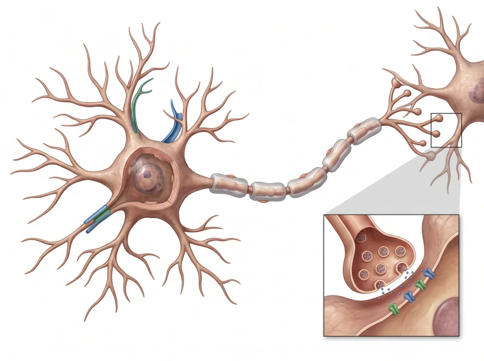
1.Dendrites
Branching extensions receive information.
2.Cell body
The cell body maintains the cell and integrates incoming signals.
3.Axon
Signals travel along the axon toward its terminals.
4.Axon terminal
The axon ends in terminals that pass the signal on to the next cell.
The axon terminal inset is magnified. The axon carries signals toward its terminals.
Central, peripheral, and the direction of traffic
The (CNS) is the brain and the spinal cord. Everything outside them belongs to the (PNS): the nerves, plus small clusters of cell bodies called ganglia.
Twelve pairs of leave the brain directly. Among them, the optic nerve carries sight, the facial nerve moves the face, and the vagus nerve descends into the chest and abdomen. Thirty-one pairs of spinal nerves leave the spinal cord and exit between the vertebrae.
Signals in these nerves travel in two directions. Sensory fibers carry information from the skin, muscles, and organs in toward the CNS, and motor fibers carry commands out to muscles and glands. Every spinal nerve carries both kinds, so a pinched spinal nerve can cause numbness, weakness, or both in the part of the body it serves.
Skeletal muscles answer to the somatic division, which you command on purpose. Heart muscle, the smooth muscle of the vessels and gut, and the glands answer to the division, which runs without your attention and has two opposing branches.
The branch prepares the body for action, with a faster heart, wider airways, blood sent to the muscles, and sweating palms. The branch settles the body for rest and digestion, with a slower heart and an active gut.
The brain, region by region
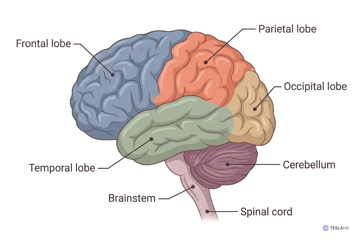
The large, folded forms the most superior part of the brain and is divided into right and left hemispheres. Each hemisphere mostly controls the opposite, or , side of the body, so a stroke in the left cerebrum weakens the right arm.
Each hemisphere has four lobes, named for the skull bones that cover them. The , the most anterior, handles planning, personality, speech, and voluntary movement. The , posterior to the frontal lobe, processes touch and body position. The , on the lateral side just superior to the ear, handles hearing and memory. The , the most posterior, processes vision.
Three structures lie deep inside the cerebrum. The is a thick band of white matter that joins the two hemispheres. The , inferior to the corpus callosum, relays nearly all sensory information up to the cortex. The , clusters of gray matter lateral to the thalamus, help start and smooth movement.
The , or "little brain," lies inferior to the occipital lobes of the cerebrum. It smooths and times the movements that the cerebrum starts, and it keeps the body balanced.
The lies anterior to the cerebellum and joins the cerebrum to the spinal cord. From superior to inferior, its three parts are the , the , and the . The medulla sets the rhythm of breathing and adjusts heart rate and blood pressure. Most cranial nerves arise from the brainstem.
On a midline sagittal image of the head, the corpus callosum is the curved band of white matter arching across the middle of the brain. The cerebellum sits posteriorly, inferior to the occipital lobe, and the brainstem descends anterior to the cerebellum into the spinal cord.
Interactive model
The brain is highlighted from the patient's left. Turn the model to see it from the side and from behind, where the cerebellum tucks under the back of the cerebrum.
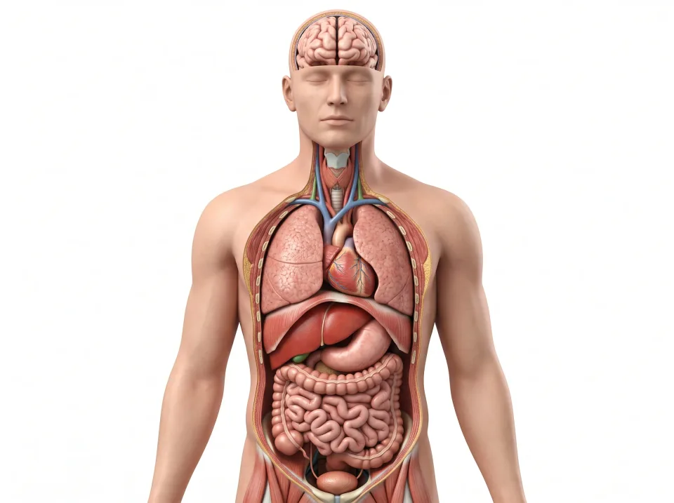
Body location map
Highlighted structure
Brain
Where: Inside the cranial cavity
Job: Processes information and coordinates many body functions.
Drag to turn. Pinch to zoom. Select a visible structure to learn about it.
Where it sits on a slice
Axial through the brain
Seen from the feet, so the patient's right is on your left.
Coverings and fluid
Three membranes, the , wrap the brain and spinal cord like the skins of an onion. The tough outer lines the inner surface of the skull. Deep to the dura lies the thin, web-like . The delicate clings to every fold of the brain's surface.
Between the arachnoid and the pia lies the , filled with (CSF), a clear liquid that floats the brain. Because the brain floats, its weight does not crush its own base, and the fluid cushions it against a knock to the head.
CSF is made inside four hollow chambers deep in the brain, the . The two lateral ventricles lie one in each hemisphere, the narrow third ventricle in the midline, and the fourth ventricle anterior to the cerebellum. Every brain scan shows the ventricles and the CSF around the brain.
CSF flows out of the fourth ventricle into the subarachnoid space, bathes the brain and cord, and is absorbed back into the blood. The whole volume, about 150 mL, is replaced several times a day. A lumbar puncture samples CSF from the subarachnoid space inferior to the end of the cord, where there is no cord to hit.
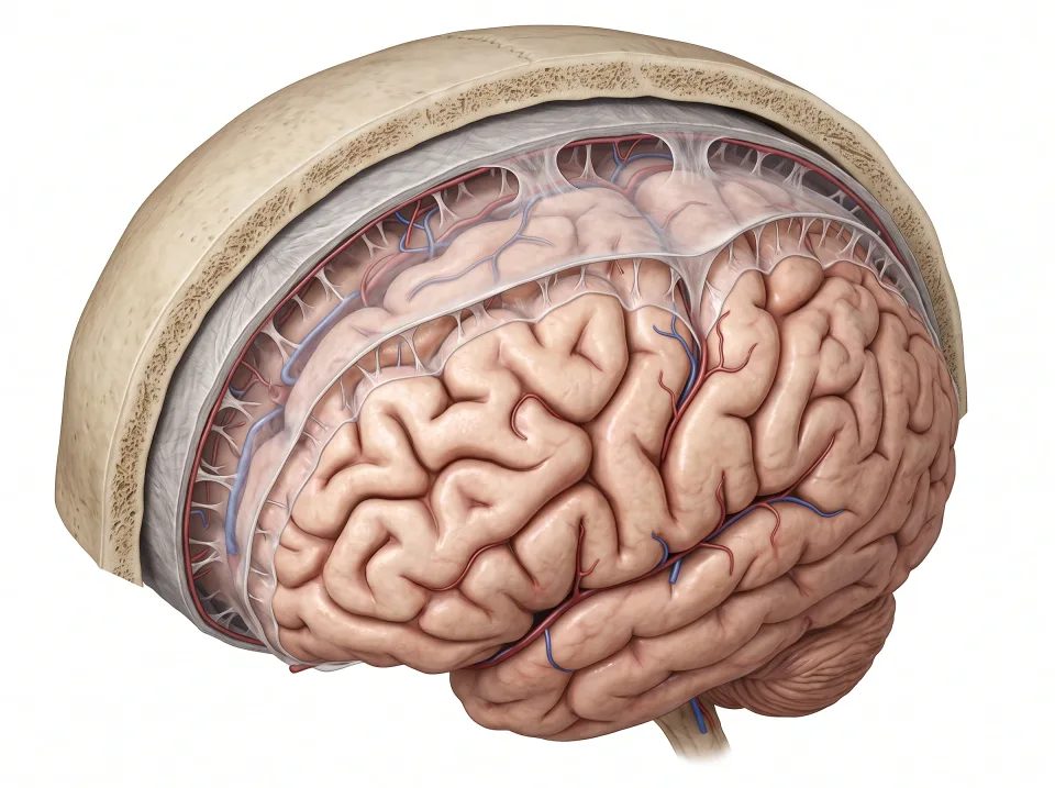
1.Dura
The tough outer covering lies beneath the skull.
2.Arachnoid and CSF
The arachnoid lies beneath the dura. CSF fills the subarachnoid space below it.
3.Pia
The delicate inner covering follows the brain surface.
Layers and spaces are enlarged for visibility. CSF lies beneath the arachnoid, not between the skull and dura.
The spinal cord and the cauda equina
The spinal cord is nervous tissue about as thick as your little finger. It runs inside the vertebral canal, the bony channel formed by the stacked vertebrae. The cord carries every message between the brain and the body inferior to the neck, and it runs the reflexes of the trunk and limbs.
During childhood the cord grows more slowly than the vertebral column. As a result, the adult cord ends near the L1 to L2 vertebral level, in a tapered tip called the . The vertebral canal continues inferiorly into the sacrum, but the cord does not.
Inferior to the conus, the canal holds a bundle of nerve roots called the , named for its resemblance to a horse's tail. Each root descends to its own opening between the vertebrae and leaves the spine there as a spinal nerve.
The cord is one continuous nervous structure, while the cauda equina is a bundle of separate roots floating in CSF. On a sagittal image of the lumbar spine, the cord tapers to the conus, and the roots continue inferiorly inside the fluid-filled canal.
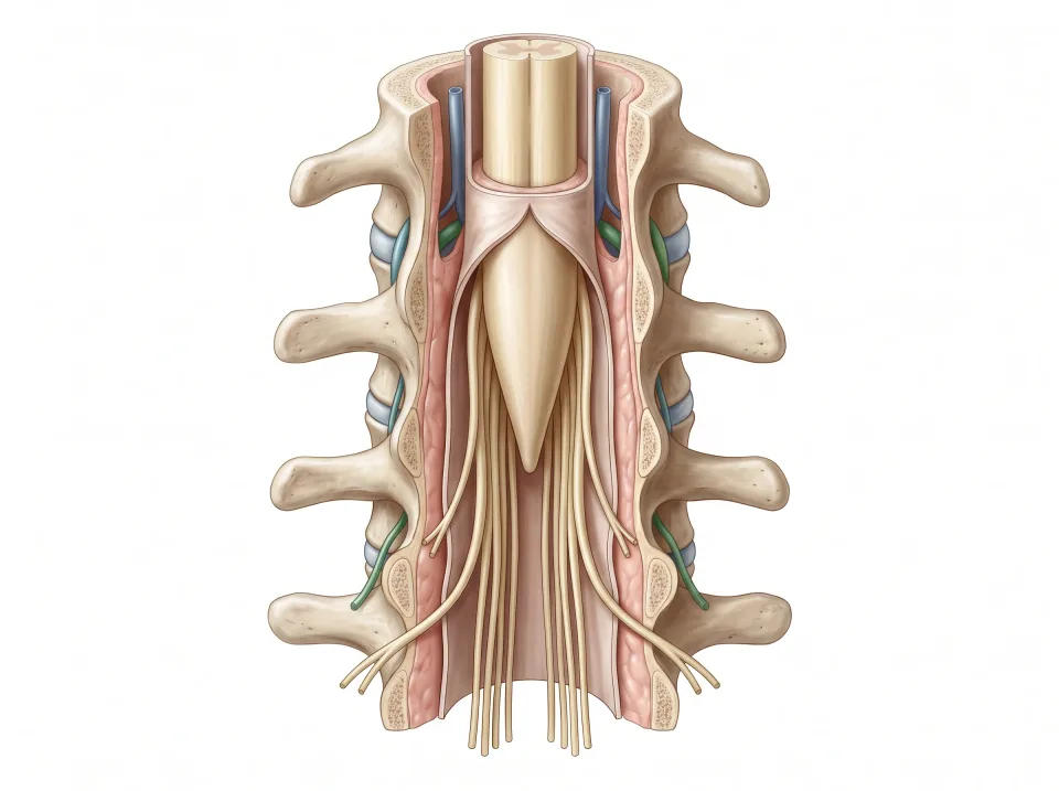
1.Spinal cord
Nervous tissue extends from the brainstem down through the vertebral canal.
2.Conus medullaris
The adult cord tapers to an end near the L1–L2 vertebral level.
3.Cauda equina
Separate nerve roots continue below the cord toward their exits from the lower spine.
Hormones and the glands that make them
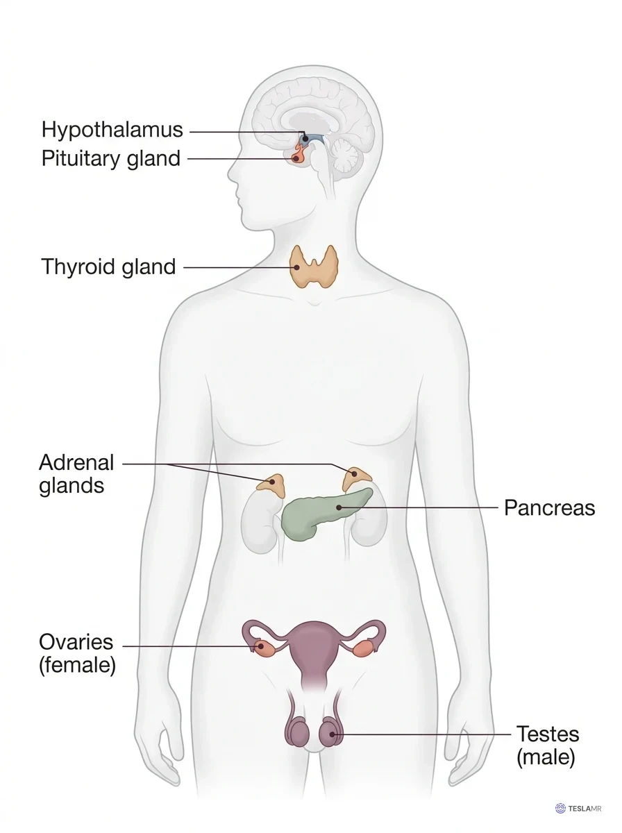
An gland has no duct, so it releases its product, a , directly into the blood, which carries it to the whole body. The figure below maps the major glands from superior to inferior.
Many glands take their orders from the , a small region at the base of the brain that is brain tissue yet makes hormones. It directs the pea-sized gland, which hangs inferior to it on a thin stalk. The pituitary rests in the ("Turkish saddle"), a saddle-shaped hollow in the sphenoid bone. Its hormones drive the , the adrenals, and the , and it also releases growth hormone. Because it drives so many glands, it is called the master gland.
In the neck, the butterfly-shaped lies anterior to the trachea, just inferior to the larynx. Its hormones set the body's metabolic rate, the speed at which cells burn fuel. Posterior to the thyroid lie four rice-grain glands, which raise blood calcium when it falls.
In the abdomen, an gland caps the superior end of each kidney and makes stress hormones such as adrenaline. Its other name, suprarenal, means "above the kidney."
The pancreas lies posterior to the stomach, and clusters of cells within it, the , release and . Insulin lowers blood glucose after a meal, and glucagon raises it between meals.
The most inferior glands on the map are the , the ovaries and testes, which make eggs or sperm along with estrogen and progesterone or testosterone.
On an axial slice just superior to the kidneys, each adrenal gland is a thin shape like an upside-down Y or V. The right adrenal lies posterior to the inferior vena cava, and the left adrenal lies lateral to the aorta.
Interactive model
The pancreas is highlighted, posterior to the stomach. Choose the adrenal glands in the list to find them superior to the kidneys.
Body location map
Highlighted structure
Pancreas
Where: Posterior to the stomach
Job: Produces digestive secretions and blood-glucose-regulating hormones.
Drag to turn. Pinch to zoom. Select a visible structure to learn about it.
Where it sits on a slice
Axial through the pancreas
Seen from the feet, so the patient's right is on your left.
The head and neck as a region
The brain fills the cranial cavity, wrapped in its and floated in cerebrospinal fluid. The two are bony sockets inferior to the frontal lobes, and each holds an eyeball, the muscles that aim it, and the optic nerve. The are air spaces in the facial bones around the nose. They are lined like the nasal cavity, and a cold can fill them with fluid.
The rests in the at the center of the skull base. It lies posterior to the sinuses and just inferior to the point where the optic nerves cross. A pituitary tumor that grows superiorly can therefore press on these nerves and narrow vision.
The neck joins the head to the chest, and every axial section through the lower neck shows its contents in the same order. In the midline, from anterior to posterior, lie the , the , and the seven vertebrae with the spinal cord inside them. The lies anterior to the trachea and wraps around it, just inferior to the .
On each side of the neck, a carries blood superiorly to the brain. An , lateral to the artery, carries blood from the brain back toward the heart, and chains of lie along both vessels.
The at the superior end of the neck include the parotid gland, anterior to each ear, and the submandibular gland, inferior to the jaw.
Key ideas to remember
- The brain and spinal cord form the , and the nerves outside them are peripheral. insulates axons and makes white matter pale. The branch readies the body for action, and the branch settles it for rest.
- From superficial to deep, the are the dura, arachnoid, and pia. is made in the four ventricles and fills the subarachnoid space around the brain and cord.
- The adult spinal cord ends near L1-L2. Inferior to that level, the vertebral canal holds the nerve roots of the .
- Hormones travel in the blood to the whole body. The directs the pituitary, which hangs inferior to it in the sella turcica.
Check what you learned
Twenty-five questions from this chapter, two of them illustrated, each with the answer, the reasoning, and a memory aid.
1 of 4: A signal starts. Dendrites carry incoming signals toward the cell body, and the neuron sends its own signal away along the axon.