Pelvic and reproductive anatomy
The pelvis contains the bladder, the rectum, and the internal reproductive organs. Learn their jobs and their anterior-to-posterior order in both the female and the male pelvis. The breast, which responds to the same hormones as the uterus, completes the reproductive picture from the chest wall.
By the end of this chapter you can
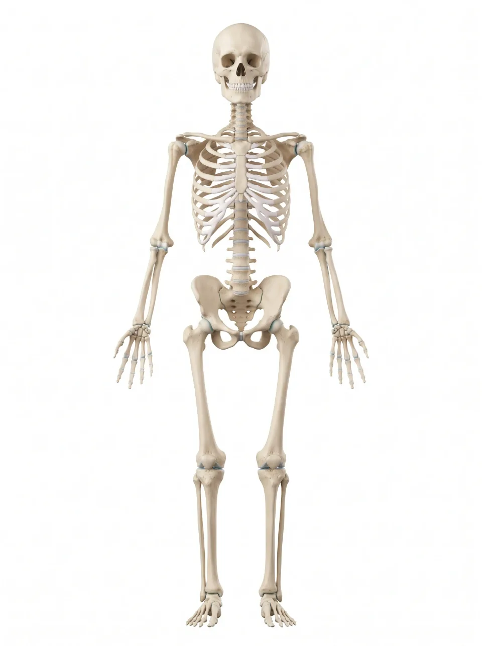
- Name the bones of the pelvis and locate the pubic symphysis, sacroiliac joints, and acetabulum on the model.
- Describe what the pelvic floor holds up, which passages open through it, and what happens when it weakens.
- Describe the anterior-to-posterior order of the bladder, reproductive organs, and rectum in a female and a male pelvis.
- Identify the parts and wall layers of the uterus and explain what the endometrium does across a menstrual cycle.
- Locate the prostate and seminal vesicles, trace sperm from the testis to the urethra, and compare the female and male urethra.
A basin of bone
The bony pelvis is a ring made of the two hip bones, the posteriorly, and the at the inferior tip of the sacrum. Shaped like a basin, it passes body weight from the spine into the lower limbs and shields the bladder, rectum, and reproductive organs inside its walls.
Each hip bone begins as three separate bones: the is superior, the is inferior and posterior, and the is anterior. During adolescence the three fuse at the , the deep socket on the lateral surface of the hip bone that holds the head of the .
The two pubic bones meet in the anterior midline at the , a pad of cartilage that barely moves. Posteriorly, the ilium of each hip bone meets the sacrum at a . Both sacroiliac joints are nearly immovable, and they transfer the weight of the trunk to the legs. They are also a common source of low back pain.
Locate these bones on the model, then feel your own. The iliac crest, the superior rim of the ilium, is the bony ridge at your waist. The ischial tuberosity, the inferior end of the ischium, is the point you sit on. The pubic symphysis is the hard midline bone just superior to the genitals.
On a coronal image through the hips, the heads of the femurs sit in the two acetabula like balls in cups. The wings of the ilium rise superior to them and flare laterally, and the bladder sits in the midline between the two hip joints.
Interactive model
The pelvic bones are highlighted. Turn the model to see the two hip bones join the sacrum at the back and meet each other at the pubic symphysis in front.
Body location map
Highlighted structure
Pelvic bones
Where: Base of the trunk, between the abdomen and thighs
Job: Supports the trunk and pelvic organs and transfers weight to the legs.
Drag to turn. Pinch to zoom. Select a visible structure to learn about it.
Where it sits on a slice
Coronal through the pelvic bones
Seen from the front, so the patient's right is on your left.
The pelvic floor
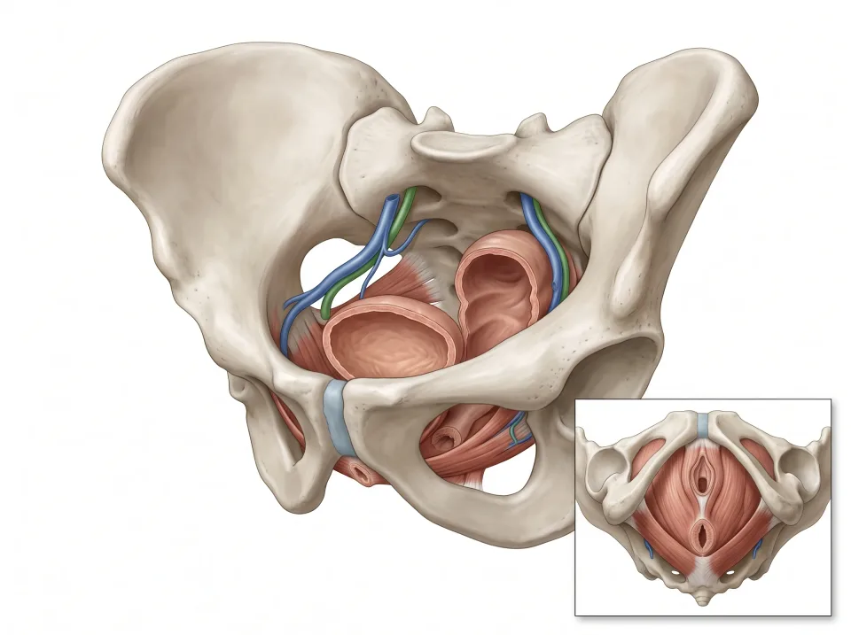
The is the sheet of muscle that forms the bottom of the pelvis, inferior to the bladder, rectum, and reproductive organs. Its main muscle is the levator ani, and the smaller coccygeus lies posterior to it. Together these muscles stretch like a hammock from the pubis anteriorly to the coccyx posteriorly, and they are also called the pelvic diaphragm.
The pelvic floor holds the bladder, rectum, and reproductive organs up against gravity. It also braces them against the pressure of coughing, lifting, and laughing.
The urethra and the anal canal pass through openings in the pelvic floor, and in women the vagina does too. These openings are where the pelvic organs reach the outside. When the floor is strong, the same muscles that lift the organs also squeeze these openings shut. This is why the levator ani matters for continence, the ability to hold urine and stool until you choose to release them.
The openings are also the weak points of the floor. Childbirth, chronic straining, and age can stretch the floor so that the bladder or uterus sags inferiorly, a condition called .
1.Organs
The bladder and rectum, with reproductive organs, sit above the pelvic floor.
2.Muscular floor
Muscles and connective tissue support the organs while allowing outlets to pass through.
The inset looks from below. The muscular floor has openings for the pelvic outlets.
Bladder in front, rectum behind
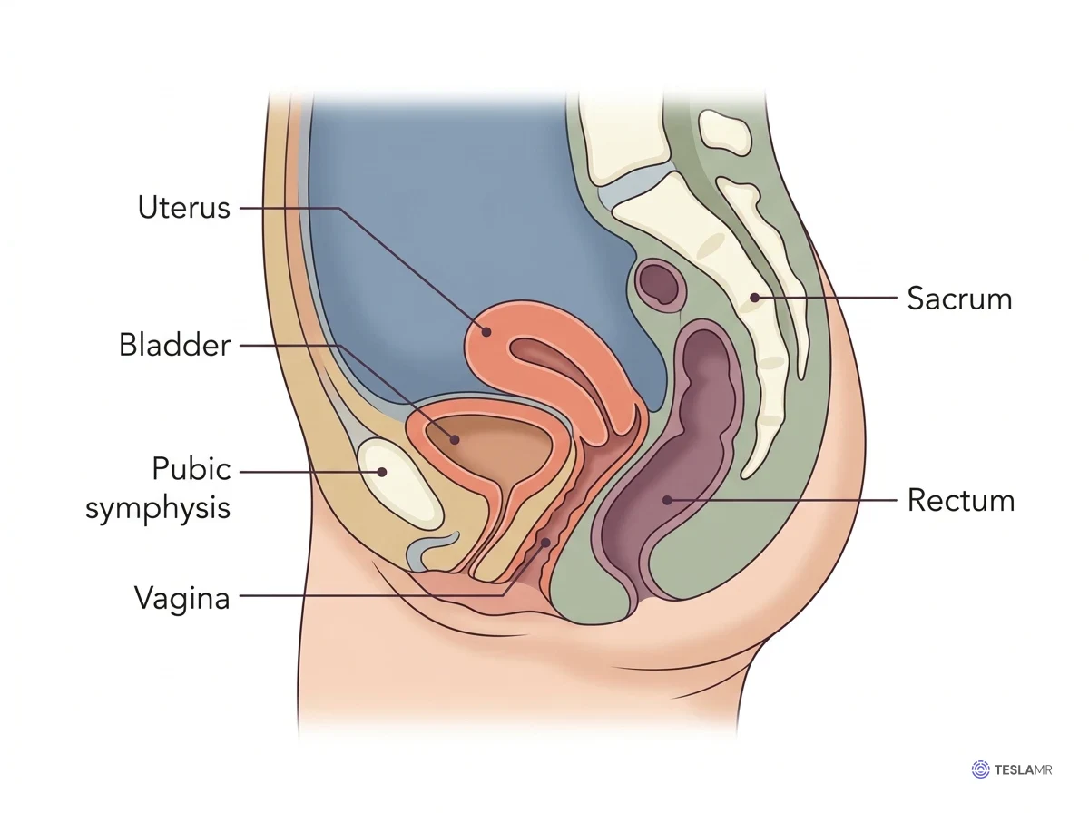
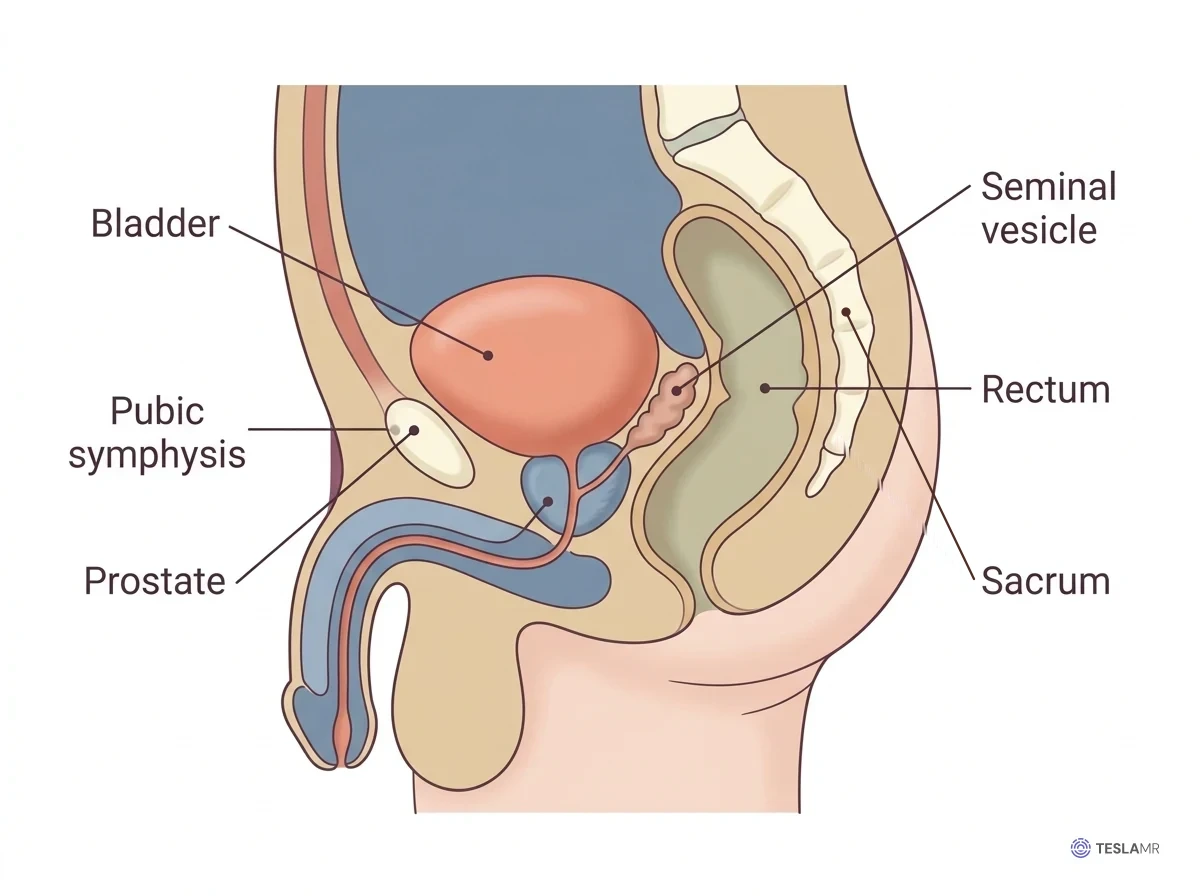
Two organs sit in every pelvis, and they anchor the whole map. The sits low and , directly posterior to the pubic symphysis and anterior to the uterus or rectum. When empty, it is a collapsed, thick-walled sac, and as it fills with urine from the two , it rises like a balloon toward the abdomen. A full bladder can be felt superior to the pubic bone.
The is the last part of the large intestine and the organ of the pelvis. It lies directly anterior to the sacrum and coccyx, following their curve, and then turns inferiorly into the anal canal.
Everything else in the pelvis fits between these two. In the figure below, two views place a female and a male pelvis side by side, and both follow the same anterior-to-posterior order. In the female pelvis, the and vagina lie posterior to the bladder and anterior to the rectum. In the male pelvis, the and occupy the same territory. The prostate lies inferior to the bladder, and the seminal vesicles lie posterior to the bladder.
Female pelvis
Male pelvis
The female pelvis: uterus, vagina, and adnexa
The is a thick-walled, pear-shaped muscular organ about 7 to 8 cm long. Its is the rounded dome superior to where the enter, its body is the main portion, and its is the narrow inferior neck that projects into the superior end of the vagina. The cervical canal through the cervix carries menstrual blood out and lets sperm in.
In most women the uterus is , meaning it tips anteriorly over the superior surface of the bladder. Its body and fundus rest on the bladder dome, so a filling bladder lifts the uterus. In roughly one woman in five, it tips posteriorly toward the rectum instead, a normal variant called .
The , a muscular tube about 8 to 10 cm long, runs inferiorly and anteriorly from the cervix to the body surface. It lies posterior to the bladder and urethra and anterior to the rectum.
The are almond-shaped glands about 3 cm long, one lateral to the uterus on each side, near the pelvic side wall. They release oocytes (egg cells) and make estrogen and progesterone, so they are endocrine glands too.
The uterine (fallopian) tubes extend about 10 cm from the superior corners of the uterus toward each ovary. Each tube opens near its ovary without touching it. Fringed projections called fimbriae sweep the released oocyte into the tube, where fertilization happens.
Together, the ovaries, tubes, and their ligaments are the , Latin for things attached alongside. A radiology report that calls the adnexa normal is describing these structures.
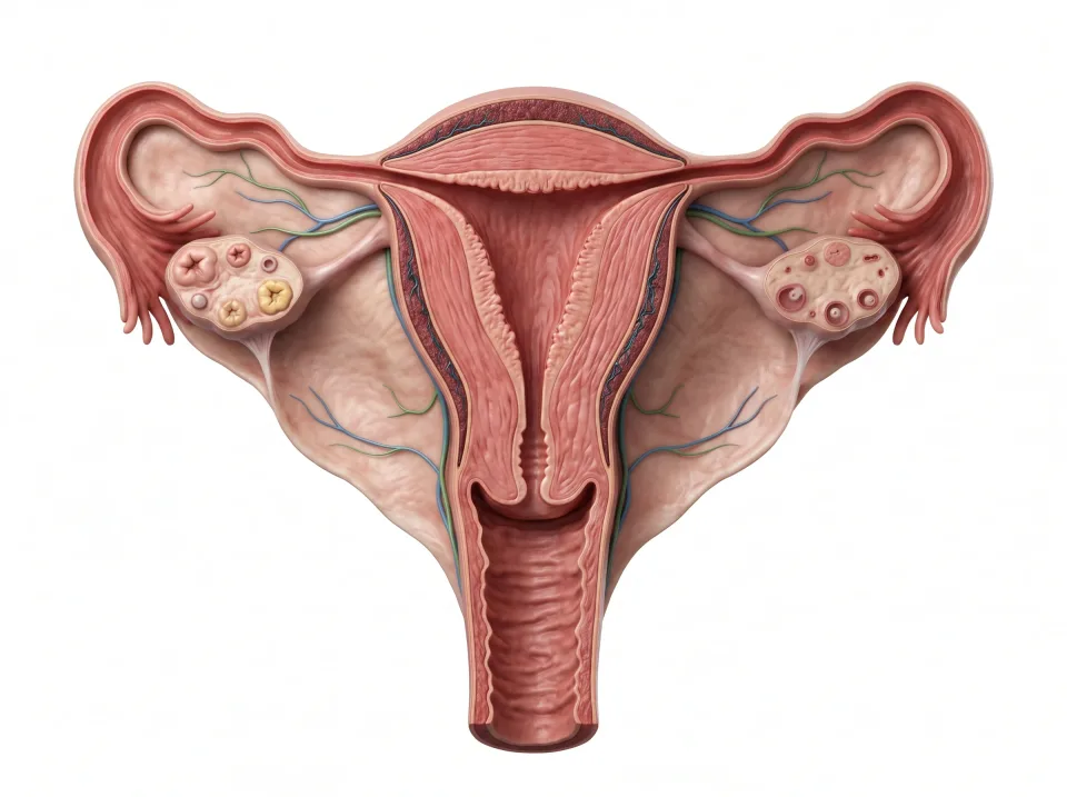
1.Ovary
An ovary releases eggs and produces hormones. One lies on each side of the uterus.
2.Uterine tube
The tube can receive an egg and carries it toward the uterus; fertilization commonly occurs in the tube.
3.Uterus
The uterus is a muscular organ where an implanted pregnancy develops.
4.Vagina
The vagina connects the cervix to the exterior, separate from the urethra.
The tubes open near the ovaries; they do not seal directly onto them.
The uterine wall, the monthly cycle, and pregnancy
From the cavity outward, the uterine wall has three layers. The is the inner lining, the is the thick muscular middle layer that contracts during childbirth, and the is the thin outer covering.
The endometrium is the layer that changes during each . In the first half of the cycle, estrogen from the ovary makes it thicken. Around the middle of the cycle, the ovary releases an egg, an event called ovulation. In the second half, progesterone keeps the lining thick and rich in blood vessels, ready for an embryo.
If no pregnancy occurs, hormone levels fall, and the thickened layer breaks down and is shed as the menstrual period, which starts the next cycle. If an embryo implants instead, the endometrium stays in place. Through the pregnancy, the uterus grows from a pear-sized organ in the pelvis into one that fills the abdomen.
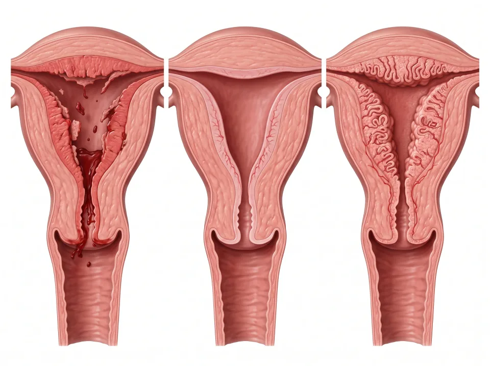
1.Menstruation
Part of the endometrium is shed if pregnancy has not occurred.
2.Lining rebuilds
Hormones stimulate growth of the lining. Ovulation occurs around the transition to the next phase, with variable timing.
3.Lining prepares
After ovulation, hormones support the lining. Without pregnancy, hormone levels fall and a new cycle begins. Cycle lengths vary.
The views compare changes in the lining, not fixed cycle days. Timing and cycle length vary.
The breast: a gland on the chest wall
The breast belongs to the reproductive system even though it lies far from the pelvis. It rests on the pectoralis major muscle of the anterior chest wall, roughly over the second to sixth ribs. Men have a small amount of breast tissue deep to the nipple.
Inside the breast, 15 to 20 lobes of milk-making glandular tissue are arranged around the nipple like the segments of an orange. Each lobe drains through a duct that opens at the nipple, and the darker ring of skin around the nipple is the areola. Fat fills the spaces between the lobes and sets most of the breast's size and shape.
The breast responds to the same hormones as the uterus. Estrogen and progesterone make it fuller in the second half of each cycle, and it grows through pregnancy. After birth, the hormone prolactin from the pituitary switches on milk production.
Most of the breast's lymph drains to the in the armpit, which is why those nodes are examined when a breast cancer is diagnosed. The upper outer quarter of the breast, its superior and lateral part nearest the armpit, holds the most glandular tissue, and most breast lumps appear there.
The male pelvis: prostate, seminal vesicles, and testes
The is a gland about the size of a walnut that sits directly inferior to the bladder and anterior to the rectum. The urethra leaves the bladder and passes straight through the middle of the prostate, and this segment is called the prostatic urethra. The posterior part of the gland, nearest the rectum, is the peripheral zone, where most prostate cancers begin. The prostate adds fluid to semen, and it tends to enlarge with age, which can squeeze the urethra and make urination slow.
The are a pair of coiled, finger-sized glands that lie posterior to the bladder and superior to the prostate. These glands contribute most of the fluid in semen.
The are the paired glands that produce sperm and the hormone testosterone. They hang outside the body cavity in the , because sperm production works best a couple of degrees below core body temperature. Sperm mature in the , a coiled tube draped over the posterior surface of each testis, before entering the vas deferens.
Each , also called the ductus deferens, is a muscular tube that carries sperm superiorly from the epididymis, through the groin, and over the bladder. Posterior to the bladder, it joins the duct of a seminal vesicle. The joined ducts enter the posterior surface of the prostate and pass through it to empty into the prostatic urethra.
On an axial slice at the level of the pubic symphysis in a male, the prostate is the rounded gland in the midline, with the pubic bone anterior to it and the rectum directly posterior to it. A few slices superior to that level, the seminal vesicles appear as a pair of small coiled glands between the bladder and the rectum.
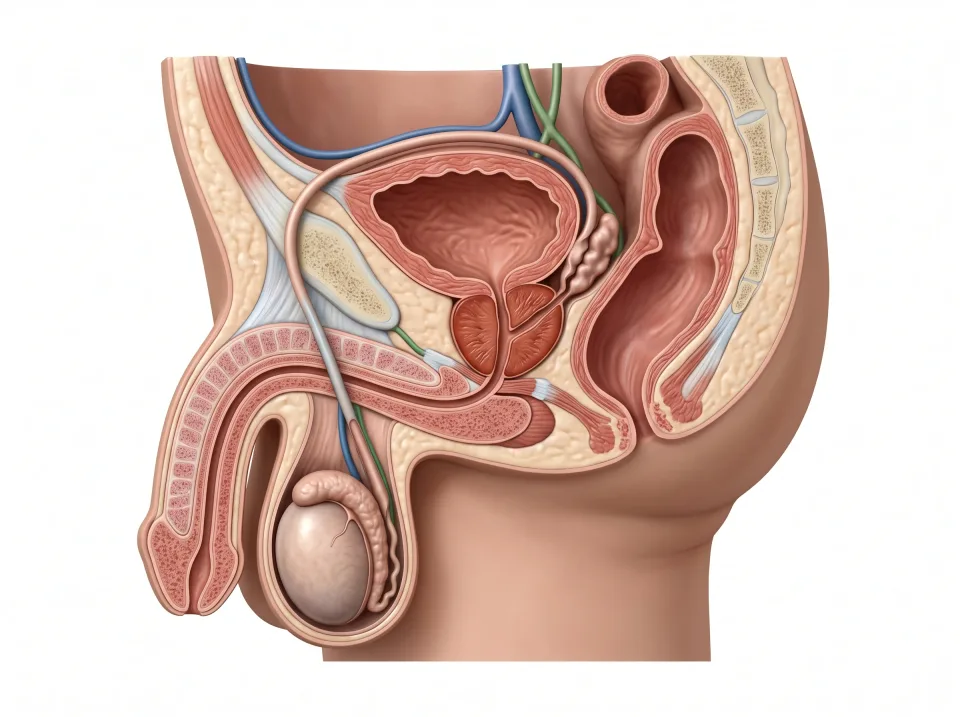
1.Testis
Sperm form in the testes, in the scrotum outside the pelvic cavity.
2.Epididymis
Sperm mature and are stored in the epididymis beside each testis.
3.Vas deferens
The vas deferens carries sperm toward the ejaculatory ducts; glands add fluid.
4.Urethra
Semen passes through the urethra to leave the body. The same tube carries urine at different times.
Two urethras of very different length
The drains the bladder in everyone, but its length and course differ sharply between the sexes. The female urethra is short, about 4 cm, and it descends almost straight from the bladder to open just anterior to the vagina.
The male urethra is about 20 cm long and passes through the prostate, then the pelvic floor, then the full length of the penis. It carries both urine and semen, though never at the same time.
The short female urethra gives bacteria a much shorter path to the bladder, so urinary tract infections are far more common in women. In men, anything that narrows the long urethra, such as an enlarged prostate, backs urine up into the bladder.
The urethra and the ureters are different tubes with similar names. Two ureters bring urine from the kidneys into the bladder, and one urethra carries it out.
Key ideas to remember
- Each hip bone is , , and fused at the acetabulum. The two hip bones meet the posteriorly at the sacroiliac joints and meet each other anteriorly at the pubic symphysis.
- In every pelvis the bladder is anterior and the rectum is posterior, with the uterus and vagina, or the prostate and seminal vesicles, between them. The pelvic floor lies inferior to all of these organs and holds them up.
- The uterus has a , body, and , and its wall is inside, in the middle, and outside. The endometrium thickens and sheds each cycle.
- The prostate lies inferior to the bladder and anterior to the rectum, and the urethra passes through it. Sperm reach that urethra by way of the epididymis, vas deferens, and seminal vesicle duct.
Check what you learned
Twenty-five questions from this chapter, two of them illustrated, each with the answer, the reasoning, and a memory aid.