Digestion and abdominal anatomy
The digestive system breaks food down, absorbs useful material, and passes what remains out of the body. Follow the continuous tube from the mouth to the anus, and learn how its neighboring organs contribute.
By the end of this chapter you can
- Trace the path of food from the mouth through the pharynx, esophagus, stomach, small intestine, and large intestine to the anus, and explain how peristalsis moves it and sphincters gate it.
- Name where most nutrient absorption happens and the organ that absorbed nutrients reach first through the portal vein.
- Explain what the liver, gallbladder, pancreas, and salivary glands each add to digestion, and why food never passes through them.
- Place the liver, stomach, spleen, appendix, kidneys, and pancreas in the correct abdominal quadrant, and say which lie posterior to the peritoneum.
One tube from mouth to anus
Everything you eat travels through one continuous passage, the or digestive tract. It begins at the mouth and passes through the (throat), , stomach, small intestine, and large intestine before ending at the and anus. Food in the tube is technically still outside the body until it crosses a living lining.
Relaxed and stretched out, the tube is roughly nine meters long. In a living person, muscle tone keeps it closer to five meters, and most of it lies folded and coiled in the .
The is the open space inside a hollow organ, where the food travels. The wall around it has an inner lining that absorbs and secretes, a middle layer of , and an outer covering. In a radiology report, an intraluminal finding lies in the passageway, and a wall lesion lies in the tissue around it.
The smooth muscle in the wall never fully rests: it contracts in coordinated waves called that push the contents along the tube. Each wave tightens behind a mouthful and relaxes ahead of it, like squeezing a tube of toothpaste.
A is a ring of muscle around the opening between two regions of the tube. It acts as a gate, holding contents in one region until the next is ready. Examples are the pyloric sphincter at the stomach outlet, the between the small and large intestine, and the anal sphincters at the end.
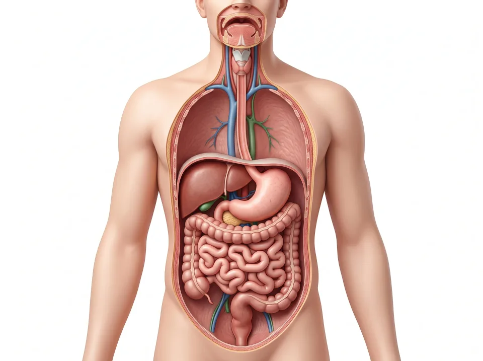
1.Mouth
Chewing and saliva begin digestion; swallowing sends food through the pharynx.
2.Esophagus
Waves of contraction move food down the chest.
3.Stomach
The stomach churns food with acid into a thick liquid.
4.Small intestine
Digestion finishes and most nutrient absorption occurs here.
5.Colon
The colon recovers water and helps form stool.
6.Rectum and anus
The rectum stores stool before it passes through the anus.
Mouth, pharynx, esophagus, and stomach
begins in the mouth, where the teeth grind food and saliva moistens it for swallowing. Saliva comes from three pairs of : the parotid anterior to each ear, the submandibular inferior to the jaw, and the sublingual inferior to the tongue.
The tongue shapes the food into a soft ball and pushes it into the pharynx, the shared passage posterior to the mouth and nose. During the swallow, a flap called the tips over the airway, so the food enters the esophagus instead of the .
The , a muscular tube about 25 centimeters long, descends through the neck and chest, first posterior to the trachea and then posterior to the heart. It passes through the esophageal hiatus, an opening in the , and joins the stomach just inferior to it. Its only job is transport: peristalsis carries each swallow to the stomach in a few seconds.
The stomach is a J-shaped bag that lies inferior to the left dome of the diaphragm. From entrance to exit, its regions are the cardia (where the esophagus enters), the fundus (the dome superior to the cardia), the body, and the (the narrowed outlet). The stomach changes size and position as it fills and empties.
The stomach stores a meal and churns it with acid into a thick liquid called . It releases the chyme in small squirts through the pyloric sphincter into the small intestine.
Interactive model
The stomach is highlighted under the left dome of the diaphragm. Turn the model to see the liver on the patient's right and the intestines inferior to the stomach.
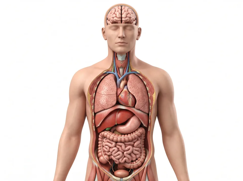
Body location map
Highlighted structure
Stomach
Where: Mainly the left upper abdomen
Job: Stores food and churns it with acid into chyme.
Drag to turn. Pinch to zoom. Select a visible structure to learn about it.
Where it sits on a slice
Axial through the stomach
Seen from the feet, so the patient's right is on your left.
The small intestine, where nutrients are absorbed
The small intestine, named for its narrow diameter, is a coiled tube about six meters long that finishes breaking food down. Most of it lies inferior to the stomach, framed by the large intestine.
The is the first and shortest of the small intestine's three segments, a C-shaped curve about 25 centimeters long that receives chyme from the stomach. The duodenum wraps around the head of the , and and pancreatic juice enter it through a shared opening.
The is the middle segment, and the is the last and longest. The ileum ends at the , where it empties into the large intestine.
The stomach churns food but absorbs very little of it. Nearly all nutrient happens in the small intestine, most of it in the duodenum and jejunum, where the broken-down food passes across the lining into the blood.
The veins that drain the intestine merge into the , which carries this nutrient-rich blood to the liver first. After the liver stores and processes what arrives, the blood leaves through the hepatic veins into the , which returns it to the heart.
The large intestine and the end of the line
The large intestine is about one and a half meters long and wider than the small intestine, which is where its name comes from. It begins in the right lower abdomen at a pouch called the , where the ileum empties in through the . The , a narrow worm-shaped tube, hangs from the cecum.
From the cecum, the frames the abdomen in four segments. The ascending colon climbs the right side to the inferior surface of the liver, where it turns and becomes the transverse colon. The transverse colon crosses the abdomen inferior to the stomach, then turns near the spleen and descends along the left side as the descending colon. Finally, the sigmoid colon makes an S-shaped bend and drops into the pelvis.
By the time material reaches the colon, digestion and nutrient absorption are essentially finished. The colon recovers water and salts, so that the liter or more of liquid arriving from the ileum becomes formed stool.
The is the straight final segment, a storage section in the pelvis anterior to the sacrum. It holds stool until it leaves through the anus, which is closed by two sphincters, one involuntary and one under your control.
Accessory organs: liver, gallbladder, pancreas
The liver, gallbladder, and pancreas are accessory organs: they deliver their products into the tube through ducts, but food never passes through them.
The liver, the largest solid organ at about 1.5 kilograms, fills most of the right upper abdomen inferior to the right dome of the . Among its many jobs, it makes , a greenish fluid that helps digest fat.
The is a small pear-shaped sac against the inferior surface of the liver. It stores bile from the liver between meals and concentrates it by pulling out water. When a fatty meal arrives, the gallbladder squeezes the stored bile into the duodenum.
Bile leaves the liver through the hepatic ducts, which unite as the common hepatic duct. The gallbladder's cystic duct joins the common hepatic duct to form the common bile duct, which descends to the duodenum. After a gallbladder is removed, bile still reaches the intestine, but it trickles in continuously instead of arriving in a burst.
The lies across the upper abdomen posterior to the stomach, with its head in the C of the duodenum and its tail reaching toward the spleen. Most of the pancreas makes digestive enzymes, which travel through the pancreatic duct to meet the common bile duct at the duodenum. It also holds the , which release and into the blood.
On an axial slice at about the level of the first and second lumbar vertebrae, the pancreas lies across the posterior part of the abdomen like a comma: its head in the curve of the duodenum on the patient's right, its body arching anterior to the aorta, and its tail reaching toward the spleen, with the splenic vein along its posterior edge. It rises toward the spleen at a slant, so a coronal image catches only part of it at a time, and a sagittal image near the midline cuts across its body between the stomach and the aorta.
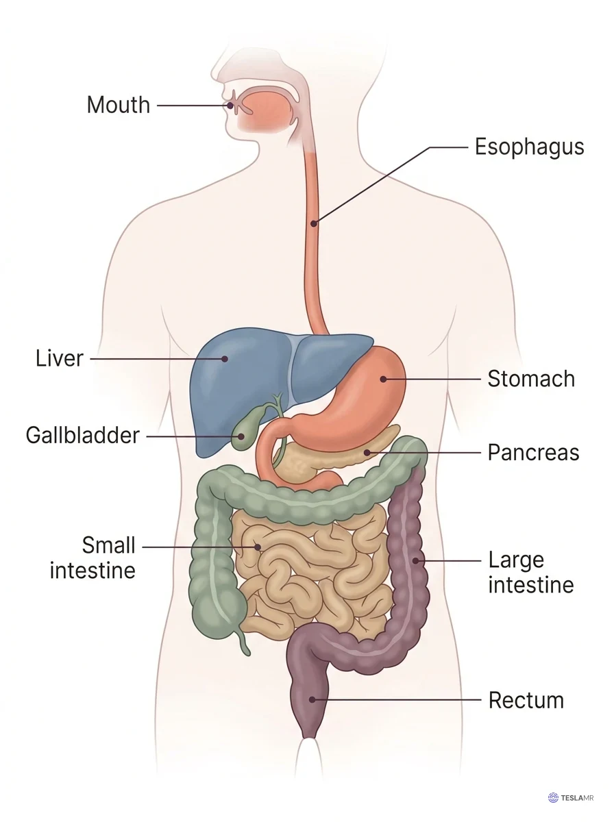
Interactive model
The liver is highlighted under the right dome of the diaphragm. Choose the pancreas in the list to find it posterior to the stomach.
Body location map
Highlighted structure
Liver
Where: Mainly the right upper abdomen
Job: Processes nutrients, produces bile, and performs many metabolic functions.
Drag to turn. Pinch to zoom. Select a visible structure to learn about it.
Where it sits on a slice
Axial through the liver
Seen from the feet, so the patient's right is on your left.
Mapping the abdomen: quadrants, regions, and the peritoneum
A vertical line and a horizontal line crossing at the navel divide the abdominal surface into four . They are named from the patient's point of view, so the right upper quadrant is on the patient's right.
The liver and gallbladder dominate the right upper quadrant, and the stomach and spleen sit in the left upper quadrant. The pancreas stretches across both upper quadrants, posterior to the stomach. The cecum and appendix lie in the right lower quadrant, where appendicitis pain settles, and the descending and sigmoid colon occupy the left lower quadrant.
A tic-tac-toe grid divides the abdomen into nine regions, centered on the umbilical region around the navel. Superior to the umbilical region lies the epigastric region, which overlies the stomach, and inferior to it lies the hypogastric or pubic region. On each side, from superior to inferior, lie the hypochondriac (covered by the ribs), lumbar or flank, and iliac or inguinal regions.
On requisitions, epigastric pain points to the stomach and pancreas, and a left flank mass to the left kidney or descending colon.
The is a thin, slippery membrane that lines the abdominal cavity and wraps most of the organs so they slide against each other. Organs suspended inside the peritoneal sac, such as the stomach, jejunum, ileum, and transverse colon, are intraperitoneal and free to shift.
Organs posterior to the peritoneum, against the posterior abdominal wall, are . They include the kidneys, the , most of the pancreas, the , and the .
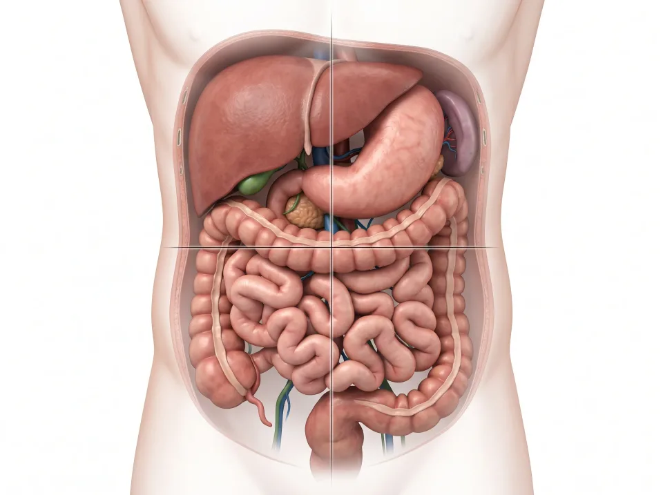
1.Right upper
Much of the liver and the gallbladder are here.
2.Left upper
The stomach and spleen lie mainly here.
3.Right lower
The cecum and usually the appendix lie here.
4.Left lower
The descending and sigmoid colon occupy this side. Organs can cross boundaries.
Right is on the viewer’s left. Organs can extend across more than one quadrant.
Key ideas to remember
- Food travels one continuous tube: mouth, pharynx, esophagus, stomach, duodenum, jejunum, ileum, cecum, colon, rectum, anus.
- Nearly all nutrients are absorbed in the small intestine, mostly the duodenum and jejunum, and the portal vein carries that blood to the liver first.
- The liver makes bile, the gallbladder stores it, and the pancreas sends digestive enzymes to the duodenum. Food never passes through any of them.
- The liver lies in the right upper quadrant, the stomach and spleen in the left upper, and the appendix in the right lower. The kidneys, pancreas, and aorta lie posterior to the peritoneum, near the spine.
Check what you learned
Twenty-five questions from this chapter, two of them illustrated, each with the answer, the reasoning, and a memory aid.
1 of 4: A mouthful enters. Food arrives in the tube. The muscular wall around it is relaxed.