Kidneys, fluid balance, and lymph
The kidneys filter the blood and turn its waste into urine. The lymphatic system returns leaked tissue fluid to the blood and filters it on the way. Learn where the organs of each system sit and the route each fluid follows.
By the end of this chapter you can
- Locate the kidneys, ureters, bladder, and urethra, and trace urine from a nephron to the outside of the body.
- Name the three regions of the kidney and the three tubes that pass through the renal hilum.
- Explain what the glomerulus filters out of the blood and what builds up in the blood when the kidneys filter less.
- Explain what lymph is, where it comes from, and where it rejoins the blood.
- Compare what the spleen filters with what a lymph node filters, and name the node groups that drain the neck, the armpit, and the groin.
Where the kidneys sit
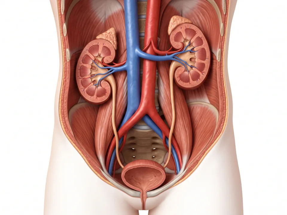
Each is a bean-shaped organ about the size of a fist, 10 to 12 centimeters long. The two kidneys lie against the posterior abdominal wall, one on each side of the spine. They reach from about the twelfth thoracic vertebra to the third lumbar vertebra, so the lower ribs shield their superior ends.
The kidneys sit posterior to the , the thin membrane that lines the abdominal cavity and wraps most of the digestive organs. Structures in this position are called , from retro, meaning behind. The aorta, the inferior vena cava, the pancreas, and the adrenal glands are retroperitoneal too.
A small adrenal gland sits like a cap on the superior end of each kidney. Because of this position, the adrenal glands are also called the suprarenal glands: supra means above, and renal means kidney.
The two kidneys are not level, and the difference is normal. The right kidney sits slightly inferior to the left because the liver, which fills much of the right upper quadrant, takes up the space superior to it.
Each kidney is packed in a cushion of fat, which lets it move with breathing. When the diaphragm descends on a breath in, it pushes the kidneys inferiorly by a centimeter or two, and they return on the breath out.
On an axial slice the kidneys lie against the posterior abdominal wall on either side of the spine. Each hilum faces anteriorly and medially toward the great vessels, with the left kidney lateral to the aorta and the right kidney lateral to the inferior vena cava. The left renal vein crosses the midline anterior to the aorta to reach the inferior vena cava.
Interactive model
The kidneys are highlighted from the back, one on each side of the spine. Turn the model to the side to see how far posterior they sit, then choose the bladder in the list to follow the ureters down to it.
Body location map
Highlighted structure
Kidneys
Where: Posterior abdomen, on either side of the spine
Job: Filter the blood and turn its waste into urine.
Drag to turn. Pinch to zoom. Select a visible structure to learn about it.
Where it sits on a slice
Axial through the kidneys
Seen from the feet, so the patient's right is on your left.
Blood in, urine out
The is a notch on the medial side of each kidney, and three tubes pass through it. The brings blood in straight from the aorta, the carries blood out to the inferior vena cava, and the carries urine away.
The kidneys receive roughly a fifth of all the blood the heart pumps. Their job is to process that blood continuously, so they take in far more blood than their own tissue needs.
A section along the long axis of a kidney shows three regions. The outer layer is the , where filtering begins. The lies deep to the cortex and is arranged in cone-shaped renal pyramids, whose tips point toward the center of the kidney.
Urine drips from the tips of the pyramids into cup-shaped , which join to form the . The renal pelvis is a funnel that collects the urine and narrows into the ureter. Urine therefore flows from the cortex through the medulla into the renal pelvis, then leaves the kidney through the hilum.
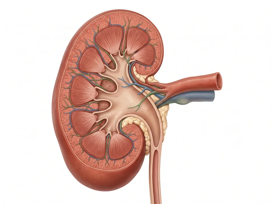
1.Cortex
The outer region is where filtering begins.
2.Medulla
The inner pyramids carry the urine inward toward the center of the kidney.
3.Renal pelvis
Urine collects into this funnel and then drains into the ureter.
How a kidney filters blood
Each kidney holds about a million , the microscopic working units that make urine. Each nephron begins with a , a tight ball of capillaries, and continues as a long, winding tube called the tubule.
is the first step in making urine, and it takes place in the glomerulus. Blood pressure pushes water out of the blood along with small dissolved substances such as salts, glucose, and a waste called urea. Blood cells and large proteins stay behind in the blood. The tubule then returns almost all of the water and the useful substances to the blood, and the rest drains toward the renal pelvis as urine.
When the kidneys fail, they filter less, so wastes such as urea build up in the blood instead of leaving in the urine. Anything else the kidneys normally clear from the blood, including some medicines, also stays in the body longer.
Ureters, bladder, and urethra
Each is a muscular tube about 25 to 30 centimeters long. From the renal pelvis, it descends along the posterior abdominal wall, staying retroperitoneal, then crosses into the pelvis and enters the posterior wall of the . Urine moves along the ureter by , the same squeezing waves of muscle that push food through the gut. Peristalsis does not depend on gravity, so urine reaches the bladder whether a person is standing or lying flat.
The bladder is a hollow muscular bag in the pelvis, directly posterior to the . Its job is to store urine, and it holds about 400 to 500 milliliters before the urge to empty becomes strong.
An empty bladder sits within the bony ring of the pelvis. As it fills, it expands superiorly into the abdomen and its wall thins. On a sagittal slice through the pelvis, a full bladder is easy to recognize: a large, smooth, fluid-filled shape just posterior to the pubic symphysis.
The is the single exit tube from the bladder to the outside of the body. Two ureters bring urine into the bladder, and one urethra carries it out.
1.Renal artery
A renal artery branches from the aorta and brings blood to a kidney.
2.Renal vein
A renal vein returns blood from a kidney to the inferior vena cava.
3.Ureter
Each ureter carries urine from a kidney to the bladder.
4.Bladder
The bladder stores urine in the pelvis.
5.Urethra
The urethra carries urine from the bladder to the outside.
Lymph: the fluid that leaks out
As blood passes through the capillaries, some of the watery plasma seeps out through their thin walls and bathes the tissue cells. Most of this fluid is drawn back into the capillaries, and the rest stays in the tissues.
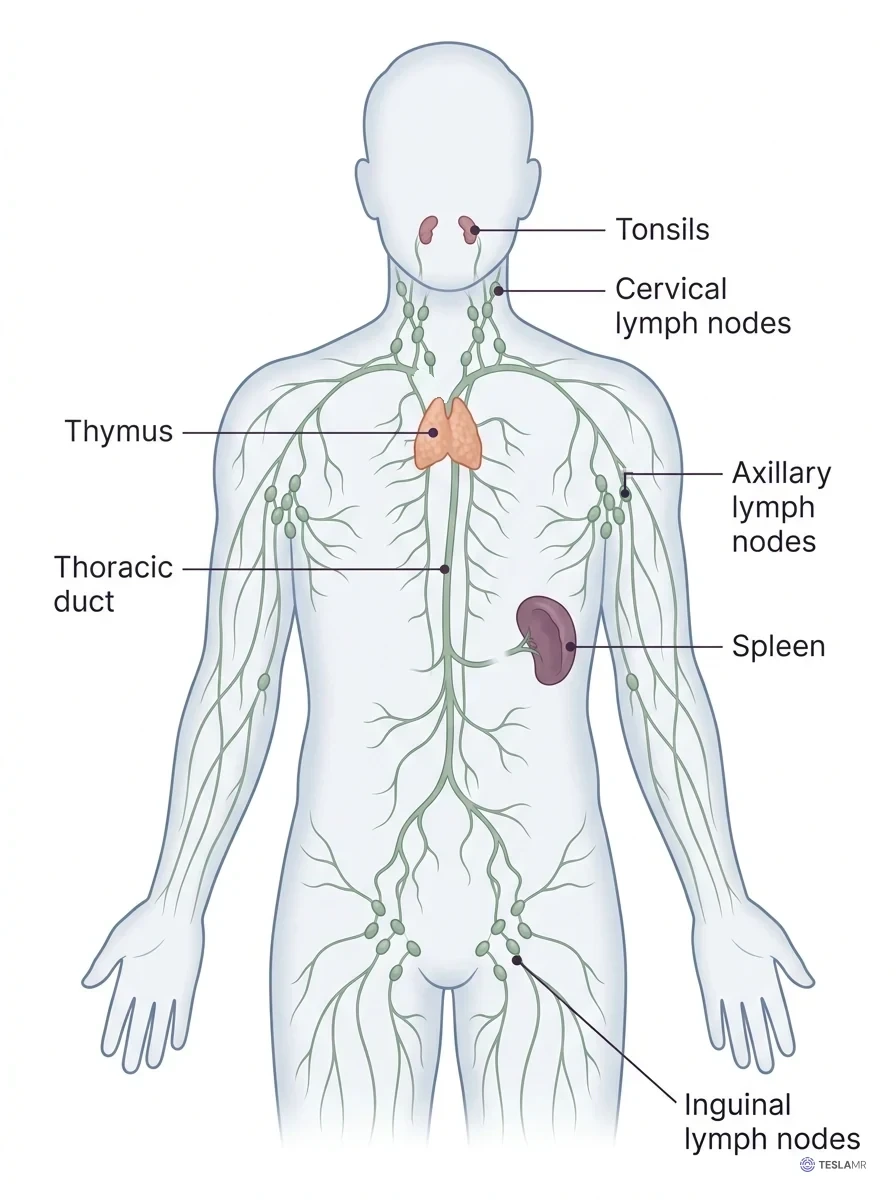
collect this leftover fluid, and once the fluid is inside them, it is called . On its way back to the blood, lymph passes through that filter it. The largest lymphatic vessel, the , ascends anterior to the spine. It empties into a large vein at the base of the left side of the neck. The fluid therefore makes a one-way trip: it leaves the blood at the capillaries and returns to the blood in the veins near the heart.
When lymph cannot drain, fluid stays in the tissue and that part of the body swells. An arm can swell this way after surgery on the armpit nodes.
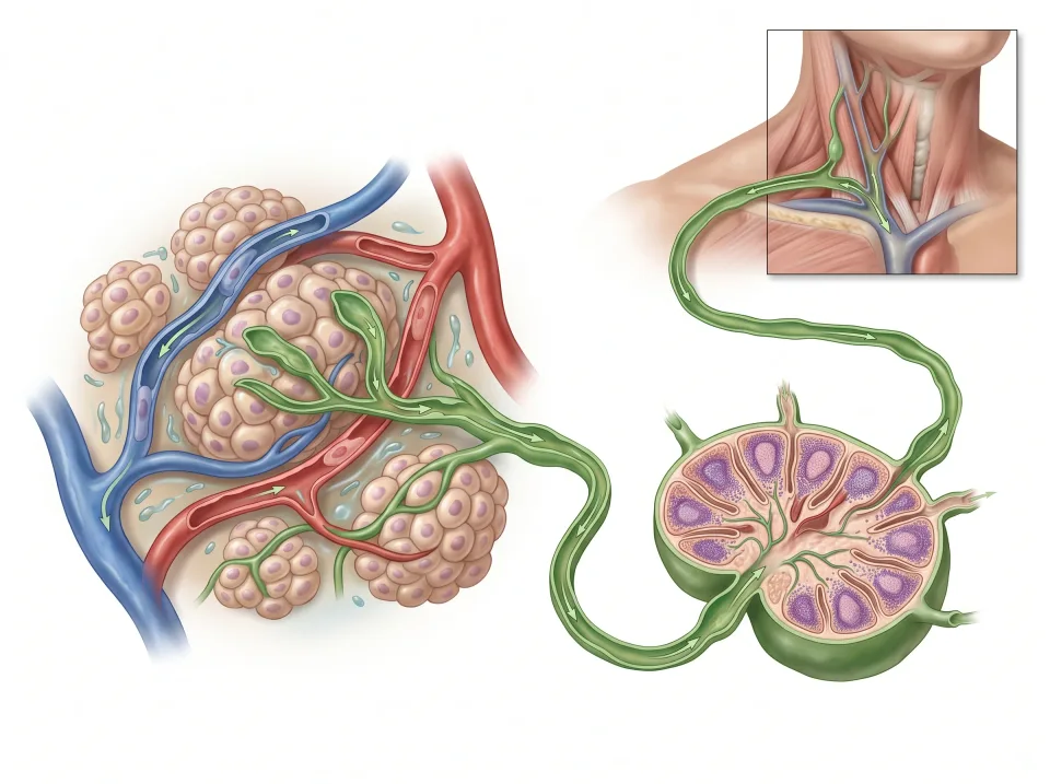
1.Tissue fluid
Fluid around cells allows exchange between blood and tissues.
2.Lymph vessel
Excess fluid enters lymphatic vessels. Valves and contractions help direct flow.
3.Node
Lymph passes through nodes, where immune cells monitor it.
4.Venous blood
Large lymphatic ducts return lymph to veins near the base of the neck.
The tissue, node and neck are shown at different scales to make the route visible.
Nodes, spleen, thymus, and tonsils
are small bean-shaped filters strung in clusters along the lymphatic vessels, and each cluster drains one region of the body. The in the neck drain the head and throat, the in the armpits drain the arm and breast, and the in the groin crease drain the leg.
Inside each node, white blood cells screen the lymph for bacteria and anything else that does not belong. With a sore throat, the cervical nodes can swell and feel tender because the white blood cells inside them multiply to fight the infection.
The is a lymphatic organ in the left upper quadrant, deep to the lower left ribs and posterior and lateral to the stomach. Blood enters the spleen through its own artery and leaves through its own vein, so the spleen filters blood, while lymph nodes filter lymph. It removes worn-out red blood cells and screens the blood for invaders.
The is a lymphatic organ posterior to the upper sternum and anterior to the heart. It helps build the immune system in childhood, then shrinks and turns to fat after puberty.
The are patches of lymphoid tissue in the throat, posterior to the mouth and nasal cavity. From there they sample whatever is swallowed or breathed in.
On an axial slice of the upper abdomen, the spleen is the crescent tucked against the lower left ribs, posterior and lateral to the stomach. On a slice through a child's chest, the thymus shows as soft tissue anterior to the great vessels and posterior to the sternum. In an adult, that space holds mostly fat.
Interactive model
The spleen is highlighted under the lower left ribs. Turn the model to see the stomach beside it, toward the midline.
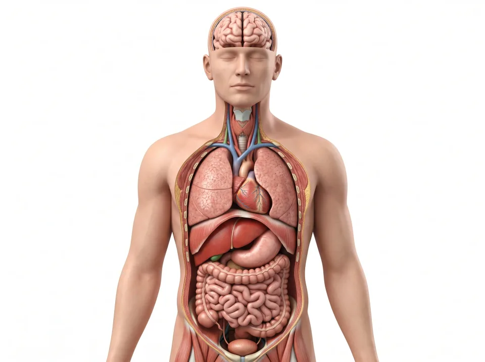
Body location map
Highlighted structure
Spleen
Where: Left upper abdomen
Job: Filters blood and supports immune responses.
Drag to turn. Pinch to zoom. Select a visible structure to learn about it.
Where it sits on a slice
Axial through the spleen
Seen from the feet, so the patient's right is on your left.
Key ideas to remember
- The kidneys lie on either side of the spine, the right slightly inferior to the left because the liver takes up the space superior to it. Each kidney filters blood through about a million .
- Blood pressure pushes water and wastes out of the blood at the , the tubule takes most of the water back, and what is left is urine. Failing kidneys filter less, so wastes build up in the blood.
- Urine travels from kidney to ureter to to : two ureters in, one urethra out.
- is leaked tissue fluid returning to the veins through the , filtered by lymph nodes along the way. The filters blood instead.
Check what you learned
Twenty-five questions from this chapter, two of them illustrated, each with the answer, the reasoning, and a memory aid.