Breathing and the lungs
Breathing brings air to the lungs, where oxygen enters the blood and carbon dioxide leaves it. Follow the airways from the nose to the alveoli, then see how the diaphragm moves the chest.
By the end of this chapter you can
- Trace the route air takes from the nose or mouth to an alveolus, naming each structure in order, and explain how the epiglottis keeps food out of the airway.
- Describe the lobes of each lung, the two layers of pleura, and the mediastinum between the lungs.
- Explain a quiet breath in terms of the diaphragm and chest volume, name the organs the diaphragm pushes down, and say why exhaling at rest takes no muscular effort.
- Describe how oxygen crosses the alveolar wall by diffusion and what carries it in the blood.
- Name the muscles that move air in and state the resting breathing rate.
The route air takes
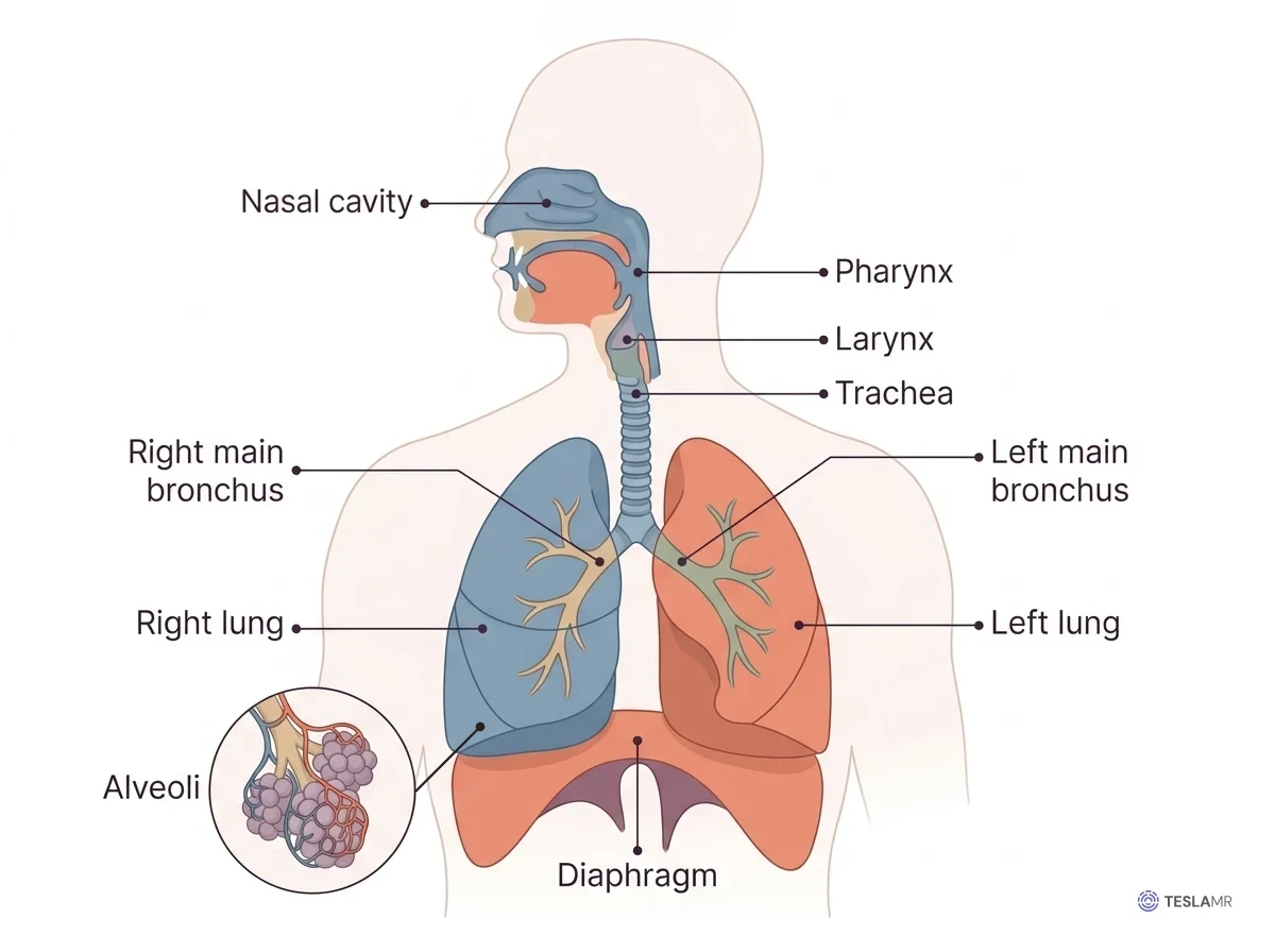
Air enters through the nose or mouth, and the nose warms, moistens, and filters it on the way in. The air then passes through the (the throat) and into the (the voice box). The cartilage of the larynx forms the bump you can feel on the anterior surface of the neck.
The begins just inferior to the larynx and carries air into the chest. It is a tube about 11 centimeters long, held open by C-shaped rings of cartilage. You can feel these rings as ridges just superior to the notch of the sternum.
At its inferior end, the trachea splits at a ridge called the into a right and a left main . The right main bronchus is wider and steeper than the left, which is why an inhaled peanut usually ends up in the right lung. Inside its lung, each bronchus branches about twenty more times, ending in no wider than a hair.
The last bronchioles open into some 300 million , tiny air sacs wrapped in capillaries. These sacs are the only part of the lung where air and blood come close enough for oxygen and carbon dioxide to cross. Every airway before them simply moves, warms, and filters the air.
On a coronal image of the chest, the trachea is a dark column of air in the midline. It forks at the carina into the two main bronchi, and the right one runs steeper than the left. Air shows as dark on MRI, so the airways and lungs always stand out against the soft tissue around them.
Where the air route and the food route part ways
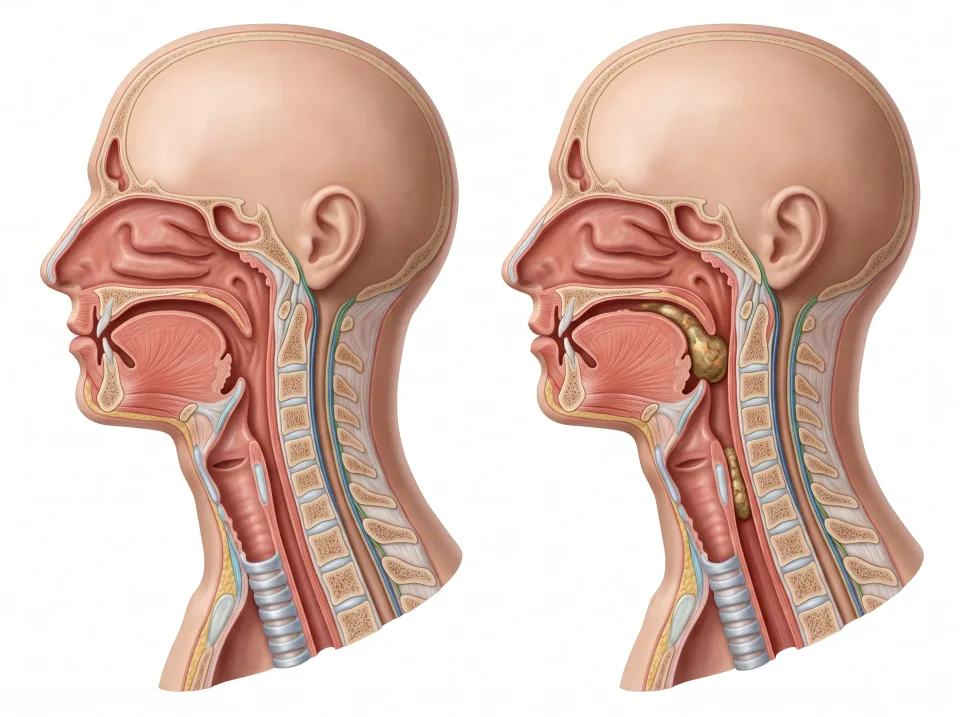
Air and food share a single passage, the , and the two routes separate just inferior to it. Air takes the anterior route, through the larynx and into the trachea. Food and drink take the posterior route, into the , a muscular tube that carries them to the stomach. In the neck, the trachea lies anterior and the esophagus lies directly posterior to it. This relationship helps you tell the airway from the food pathway.
A leaf-shaped flap of cartilage called the keeps food out of the airway. During breathing it stands upright at the superior end of the larynx, leaving the airway open. When you swallow, the larynx rises and the epiglottis tips backward like a trapdoor to cover the opening of the larynx. At the same moment, the vocal cords close just inferior to it. The mouthful slides over the closed airway into the esophagus, and between swallows the epiglottis stands upright again so breathing can resume.
1.Air
Air travels through the larynx into the trachea, toward the lungs.
2.Food
Swallowing directs food into the esophagus, behind the trachea. Several coordinated actions protect the airway.
The trachea is in front of the esophagus. Several coordinated actions protect the airway during swallowing.
The lungs and their coverings
The is the central compartment of the chest, and the lungs lie lateral to it, one on each side. It holds the heart, the great vessels, the trachea and main bronchi, the esophagus, and the thymus.
Each lung is shaped like a cone. Its pointed apex rises just superior to the first rib and the clavicle (collarbone), and its broad base rests on the diaphragm. On its medial surface is the hilum, the doorway where the main bronchus and pulmonary vessels enter.
Deep fissures divide the right lung into three : upper, middle, and lower. The left lung has only an upper and a lower lobe. It is smaller because the heart sits slightly left of the midline and presses into it. The dent in its anterior edge where the heart fits is the .
Each lung is wrapped in the , a thin, slippery membrane with two layers. The visceral pleura clings to the lung and dips into its fissures. The parietal pleura lines the rib cage and the superior surface of the diaphragm.
Between the layers is the , which in health holds only a film of fluid. That film lets the lung glide against the chest wall. Its surface tension also holds the layers together, so the lung expands whenever the chest wall does.
Interactive model
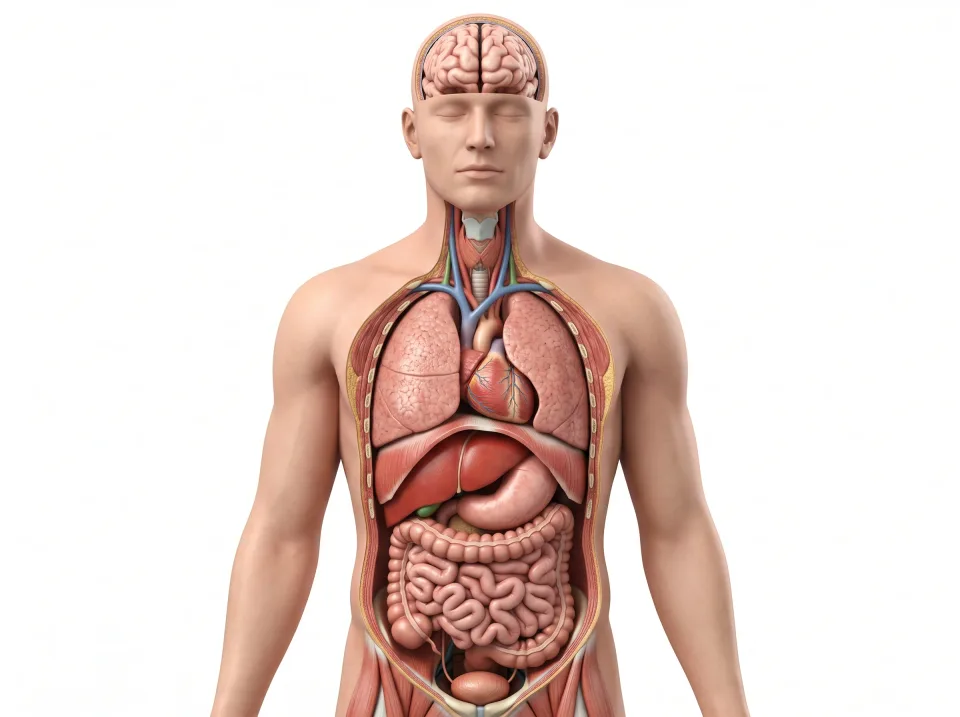
The lungs are highlighted. Turn the model to see the heart between them in the mediastinum and the base of each lung resting on the diaphragm.
Body location map
Highlighted structure
Lungs
Where: Right and left sides of the thoracic cavity
Job: Exchange oxygen and carbon dioxide with blood.
Drag to turn. Pinch to zoom. Select a visible structure to learn about it.
Where it sits on a slice
Axial through the lungs
Seen from the feet, so the patient's right is on your left.
The muscles that move air
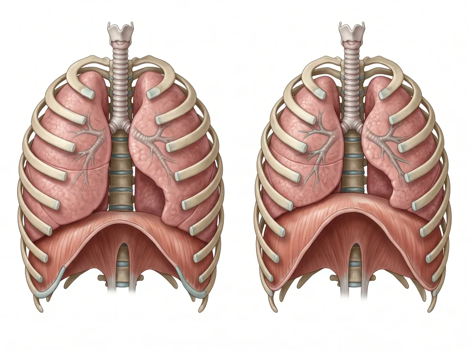
The lungs have no skeletal muscle of their own, so the muscles around them stretch and release them. The main one is the , a dome-shaped sheet of skeletal muscle that lies inferior to the lungs and separates the chest from the abdomen. When the diaphragm contracts, its dome flattens and drops toward the abdomen, and when it relaxes, the dome rises back toward the lungs.
The rim of the diaphragm attaches to the lower ribs, the sternum, and the upper lumbar spine. The aorta, the inferior vena cava, and the esophagus each pass through an opening in the diaphragm on their way between the chest and the abdomen.
The fill the spaces between the ribs. During a breath in, the external intercostals swing the ribs up and out, the way a bucket handle lifts away from the bucket. That motion makes the chest wider while the diaphragm makes it taller.
On a coronal image the diaphragm is a thin curved line, with the dark lungs superior to it and the liver and spleen inferior to it. Because the liver lies inferior to its right dome, that dome usually sits slightly superior to the left one.
Interactive model
The diaphragm is highlighted. Turn the model to the side to see its dome under the lungs, with the liver and stomach inferior to it.
Body location map
Highlighted structure
Diaphragm
Where: Between chest and abdomen
Job: Its contraction helps draw air into the lungs.
Drag to turn. Pinch to zoom. Select a visible structure to learn about it.
Where it sits on a slice
Coronal through the diaphragm
Seen from the front, so the patient's right is on your left.
What moves when you breathe
A breath in, or , begins when the diaphragm contracts and its dome descends toward the abdomen. At the same time, the external intercostals lift the ribs, and together the two actions enlarge the chest cavity. Because the pleura couples the lungs to the chest wall, the lungs stretch with it and air flows in to fill them.
The liver, spleen, stomach, and kidneys lie just inferior to the diaphragm, so they move with it. As the dome descends, it pushes these organs down a centimeter or two with a quiet breath and several with a deep one. They rise back into place as the diaphragm relaxes on the breath out.
A quiet breath out, or , takes no muscular effort. The diaphragm and intercostals relax, and the stretched lungs and chest wall spring back to their resting size like a released rubber band. This springing back, called elastic recoil, makes the chest smaller, and air flows out. Only a fast or forceful breath out needs muscles to push.
The movement of air into and out of the lungs is called . The sets its rhythm automatically, about 12 to 20 breaths a minute at rest, and each breath moves roughly half a liter of air.
Gas exchange across the alveolar wall
Once air reaches an alveolus, takes place across the alveolar wall. This wall is a single layer of flattened cells pressed against the thin wall of a pulmonary capillary. An alveolus needs two deliveries at once: fresh air on one side of its wall and flowing blood on the other.
The gases cross the wall by : with no pump involved, each one drifts from where it is concentrated to where it is scarce. Air arriving in an alveolus is rich in oxygen and poor in carbon dioxide, while blood arriving from the body is the reverse. As a result, oxygen drifts into the blood, and carbon dioxide drifts into the alveolus to be breathed out.
Every red blood cell is packed with , an iron-containing protein. It picks up oxygen in the lungs, where oxygen is plentiful, and releases it in working tissues, where oxygen is scarce. Hemoglobin carries around 98 percent of the oxygen in your blood, and its iron is what turns oxygenated blood bright red.
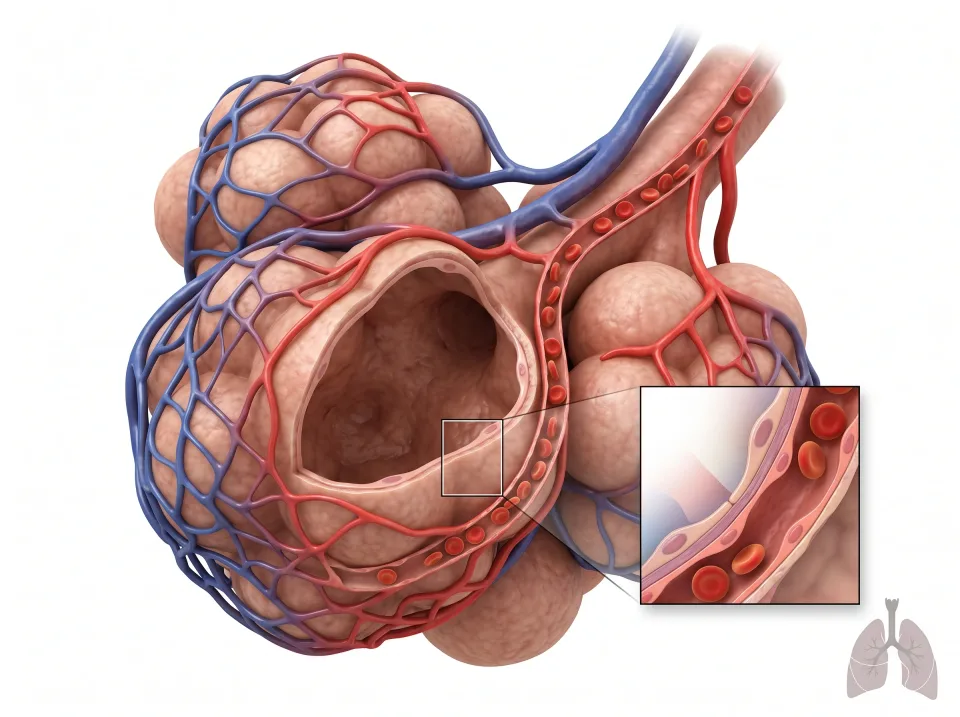
1.Oxygen enters blood
Oxygen diffuses across the thin air-sac and capillary walls into blood.
Oxygen: air space → blood
2.Carbon dioxide leaves blood
Carbon dioxide diffuses in the opposite direction and leaves the body in exhaled air.
Carbon dioxide: blood → air space
The inset enlarges the thin barrier. Oxygen moves from air to blood; carbon dioxide moves from blood to air.
Key ideas to remember
- Air travels from the nose or mouth through the pharynx, larynx, trachea, carina, main bronchi, and bronchioles to the alveoli. Only the alveoli exchange gas.
- The right lung has three lobes and the left has two, and visceral pleura covers each one. The mediastinum between the lungs holds the heart, great vessels, trachea, and esophagus.
- Breathing in is muscular: the diaphragm contracts and descends, pushing the liver, spleen, and kidneys down, while the external intercostals lift the ribs. Breathing out at rest is passive elastic recoil.
- Oxygen crosses into the blood by diffusion and rides on hemoglobin. The brainstem paces breathing at 12 to 20 breaths a minute.
Check what you learned
Twenty-five questions from this chapter, two of them illustrated, each with the answer, the reasoning, and a memory aid.
1 of 5: Breathing. The epiglottis stands upright, so air from the nose passes down the pharynx, through the larynx, and into the trachea.