The heart, blood, and circulation
The heart and blood vessels form a connected delivery system. Learn where the heart sits, how its chambers direct blood, and how blood serves the tissues.
By the end of this chapter you can
- Locate the heart in the mediastinum and describe which way the apex points.
- Name the four chambers and four valves in the order blood passes through them.
- Trace a red blood cell through the pulmonary and systemic circuits, naming the great vessels along the way.
- Explain why arteries and veins are named by direction of flow and not by oxygen content.
- List the four components of blood with the job of each, and read a blood pressure as systolic over diastolic.
Where the heart sits
The is a muscular pump, about the size of a closed fist, that sends blood carrying oxygen, nutrients, and hormones to every tissue. It sits in the , the central compartment of the chest between the two lungs. The lies anterior to the heart, the spine lies posterior to it, and the lies directly inferior to it. About two-thirds of the heart lies to the patient's left of the .
The heart is shaped like a blunt cone lying on its side. Its pointed tip, the apex, is directed inferiorly, anteriorly, and to the patient's left. Its broad base, where the great vessels attach, faces superiorly, posteriorly, and to the patient's right. With each beat the apex swings anteriorly and taps the chest wall just inferior and medial to the left nipple, at about the fifth rib space. This tap, the apex beat, is the quickest way to locate the heart on a living person.
The is a double-walled sac wrapped around the outside of the heart. A thin film of fluid between its two layers lets the beating heart slide without friction.
On an axial MRI slice through the chest, the heart is the large soft-tissue shape near the center, lying mostly on the patient's left. The dark, air-filled lungs lie lateral to it on each side.
Interactive model
The heart is highlighted between the lungs. Turn the model to the side to see the diaphragm directly inferior to it and the spine posterior to it.
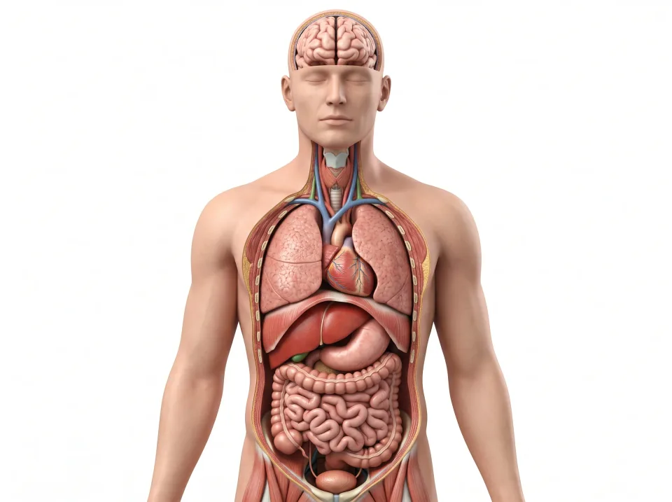
Body location map
Highlighted structure
Heart
Where: Between the lungs in the thorax
Job: Pumps blood through pulmonary and systemic circuits.
Drag to turn. Pinch to zoom. Select a visible structure to learn about it.
Where it sits on a slice
Axial through the heart
Seen from the feet, so the patient's right is on your left.
Four chambers and four valves
Inside, the heart is two pumps, a right pump and a left pump, and each has a superior receiving chamber and an inferior pumping chamber. The (one atrium, two atria) are the superior chambers, and they collect blood arriving from the . The are the inferior chambers, and they do the hard work of pushing blood out into the .
A muscular wall called the separates the right side from the left, so the two blood streams never mix in a healthy heart. The right side receives oxygen-poor blood from the body and pumps it to the lungs. The left side receives oxygen-rich blood from the lungs and pumps it to the whole body. That longer trip is why the wall of the left ventricle is about three times thicker than the wall of the right.
Four valves keep blood moving in one direction through the heart. The lies between the right atrium and the right ventricle. The , also called the bicuspid valve for its two leaflets, lies between the left atrium and the left ventricle. The guards the exit from the right ventricle, and the guards the exit from the left.
Valves have no muscle of their own, so they swing open and snap shut in response to pressure differences on either side. The "lub-dub" heard through a stethoscope is the sound of the valves closing.
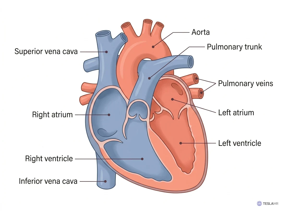
The route: two circuits joined in one pump
Blood travels through two circuits, and the heart pumps both of them. The carries blood from the right side of the heart to the lungs and back to the left side. The carries blood from the left side of the heart to every other tissue and back to the right side. Blood alternates between the two circuits, so a red cell returning from a calf muscle passes through the lungs before it can go anywhere else.
The route begins where blood returns from the body, through the superior or into the right atrium. From the right atrium, blood passes through the tricuspid valve into the right ventricle, which pumps it through the pulmonary valve into the . The trunk divides into the right and left , which carry the blood to the lungs to pick up oxygen and give up carbon dioxide.
Four return the oxygen-rich blood to the left atrium. It passes through the mitral valve into the left ventricle, and a strong contraction sends it through the aortic valve into the . From the aorta it travels out to a tissue, releases its oxygen in a bed, and returns through the veins to begin the loop again. The whole trip takes about a minute at rest.
Arteries, veins, and capillaries
Arteries and veins are named for the direction of blood flow relative to the heart. An carries blood away from the heart (A for away), and a carries blood toward it, whatever the oxygen content of that blood. This is why the pulmonary arteries, which carry oxygen-poor blood from the right ventricle to the lungs, are still arteries. Likewise, the pulmonary veins carry oxygen-rich blood from the lungs to the left atrium and are still veins.
Arteries have thick, elastic, muscular walls that withstand the pressure surge of each heartbeat. Veins have thinner walls and carry blood at low pressure. Many veins, especially in the limbs, contain one-way valves that keep blood from flowing backward and pooling. Arteries branch into smaller arterioles, which feed the capillaries, and the capillaries collect into venules, which join to form veins.
are the vessels where blood exchanges materials with the tissues, and their walls are a single layer of flattened cells. Oxygen, glucose, and hormones pass out through this thin wall into the tissue fluid, while carbon dioxide and wastes pass back into the blood. No cell in the body is far from a capillary.
is the flow of blood through the small vessels of a tissue, and it is what keeps the cells there alive. A tissue is well perfused when blood flows steadily through its capillaries.
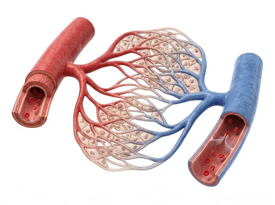
1.Artery
An artery carries blood away from the heart.
2.Capillaries
Thin capillary walls allow exchange with surrounding tissues.
3.Vein
A vein carries blood toward the heart. Many limb veins have valves.
Example of systemic circulation. Red and blue distinguish the two sides; vessel names describe direction relative to the heart.
The great vessels and the heart's own supply
The great vessels are the large vessels attached to the superior end of the heart. The leaves the left ventricle and rises as the ascending aorta. The leaves the right ventricle anterior to the aorta and divides into the right and left pulmonary arteries.
The aorta then curves posteriorly and to the patient's left as the aortic arch. It descends along the left side of the spine as the thoracic and then the abdominal aorta. Three branches leave the arch for the head and arms: the brachiocephalic trunk for the patient's right side, then the left common and the left .
The (SVC) drains the head, neck, arms, and upper chest into the superior end of the right atrium. The (IVC) drains everything inferior to the diaphragm. It ascends along the spine on the patient's right and enters the inferior end of the right atrium. On an axial slice of the upper abdomen, the IVC lies to the patient's right of the aorta.
The heart muscle, the , is too thick to be fed by the blood inside its chambers. Its supply comes from the right and left , which branch from the ascending aorta just superior to the aortic valve. They run along the surface of the heart and send branches deep into the muscle. Coronary veins drain the used blood into the right atrium. A blocked coronary artery starves a patch of myocardium, causing a heart attack.
Interactive model
The aorta is highlighted. Follow it from the heart over the arch and down the trunk, then choose the venae cavae to find the inferior vena cava on the patient's right of the aorta.
Body location map
Highlighted structure
Aorta
Where: From the heart through thorax and abdomen
Job: Carries blood from the left ventricle to the systemic arteries.
Drag to turn. Pinch to zoom. Select a visible structure to learn about it.
Where it sits on a slice
Sagittal through the aorta
Seen from the patient's left, with the face toward your left.
Vessels in the head, trunk, and limbs
In the neck, the right and left common ascend lateral to the windpipe and carry blood to the head and brain. The internal lie just lateral to the carotids and return blood from the head toward the heart.
The passes deep to the clavicle (collarbone) and becomes the axillary artery in the armpit. In the upper arm it is the , the vessel heard through a stethoscope at the anterior elbow when blood pressure is taken. At the wrist its branches are the radial artery on the lateral, thumb side and the ulnar artery on the medial, little-finger side.
In the abdomen, the aorta gives off paired to the kidneys. At about the level of the navel it divides into the right and left common , which supply the pelvis and legs.
Each common iliac artery continues as the external iliac artery and then as the , which passes through the groin into the anterior thigh. Posterior to the knee, the same vessel is called the . The leg veins carry the same names in reverse: the femoral vein becomes the external iliac vein and finally joins the IVC.
The carries blood from the stomach, intestines, spleen, and pancreas to the liver instead of straight back to the heart. The liver therefore gets first pass at most of what is absorbed from a meal. After processing, the blood leaves through the hepatic veins into the IVC.
On axial slices of the abdomen superior to about the level of the navel, the aorta is a single round vessel just anterior to the spine and slightly to the patient's left. Slices inferior to that level show two smaller circles instead: the common iliac arteries heading toward the pelvis.
What blood is made of
An adult has about five liters of blood. A little more than half of that volume is , the pale yellow liquid that carries the blood cells. Plasma is mostly water, and it also holds proteins, salts, sugar, hormones, and dissolved gases. The rest of the blood is cells, almost all of them red blood cells.
Red blood cells, which the bone marrow makes continuously, are small discs with no nucleus. Each is shaped like a doughnut whose hole did not quite punch through. They are packed with , an iron-containing protein that gives blood its red color. Hemoglobin picks up oxygen in the lungs and releases it in the tissues.
are larger than red cells but far fewer, and each one has a nucleus. As the working cells of the immune system, they patrol the body and defend it against infection.
are small fragments of cells that stick to a damaged vessel wall and start a clot, working together with clotting proteins in the plasma.
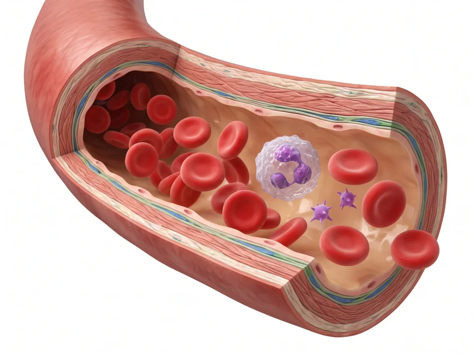
1.Red cells
Red cells contain hemoglobin, which carries oxygen.
2.White cells
White cells help defend against infection and other threats.
3.Platelets
Platelets are cell fragments that help stop bleeding.
4.Plasma
Plasma is the liquid carrying cells, proteins, nutrients, and wastes.
Cells and vessel wall are enlarged to show structure; proportions are illustrative.
Blood in motion
The beat, the pulse, and blood pressure
Each heartbeat has a contraction phase and a relaxation phase. is the contraction phase, when the ventricles eject blood into the pulmonary trunk and aorta. is the relaxation phase, when the ventricles refill with blood from the atria. At 60 beats per minute one full cycle lasts a second, and at faster rates each cycle is shorter, mostly by trimming diastole.
The heart starts each beat itself, without a signal from a nerve. The is a patch of specialized cells in the wall of the right atrium. It fires on its own about once a second and sets the pace for the whole heart, so it is called the natural pacemaker.
Every ventricular contraction sends a pressure wave along the elastic artery walls, and that wave is the pulse. It can be felt wherever an artery lies close to the skin, superficial to a bone. Examples are the radial artery at the wrist, the carotid artery in the neck, and the femoral artery at the groin.
Blood pressure is the force of blood against the artery walls, written as two numbers. A typical resting reading is about 120 over 80 millimeters of mercury. The first number is the systolic pressure, the peak pressure in the arteries during each ventricular contraction. The second is the diastolic pressure between beats, while the ventricles refill.
Key ideas to remember
- The heart sits in the mediastinum between the lungs, about the size of a fist, with its apex pointing inferiorly, anteriorly, and to the patient's left.
- Blood goes right atrium, right ventricle, lungs, left atrium, left ventricle, aorta, body, and back.
- Arteries carry blood away from the heart and veins carry it back. The pulmonary arteries are the oxygen-poor exception.
- Five liters of blood: plasma carries, red cells carry oxygen on hemoglobin, white cells defend, platelets clot.
Check what you learned
Twenty-five questions from this chapter, two of them illustrated, each with the answer, the reasoning, and a memory aid.
1 of 4: Filling. Veins return blood to both atria, and it flows on through the open tricuspid and mitral valves into the relaxed ventricles. This relaxed, refilling phase is diastole.