How the body is organized
Every structure in the body has a location and a job. Using the location words learned in the first two chapters, this chapter builds the body up from cells to tissues, organs, and systems. It then places the organs in the cavities that hold them. This is the framework that carries you through the rest of the course.
By the end of this chapter you can
- Explain the difference between anatomy and physiology using the heart as an example.
- Order the levels of organization from cell to whole person, describe how oxygen crosses tissue fluid to reach a cell, and explain negative feedback with the body's thermostat as the example.
- Name the four tissue types with an everyday example of each, and describe the epidermis, dermis, and subcutaneous layer.
- Place the brain, heart, lungs, digestive organs, and bladder in their body cavities, and name the serous membranes that line the chest and abdomen.
Two questions about every structure
is the study of the body's structures: what each one is and where it sits. is the study of what those structures do and how they work. Each structure in the body can be described both ways: by its anatomy and by its physiology.
The heart's anatomy is its structure and its position. The heart is a muscular organ about the size of your fist. It is located in the chest between the two lungs, slightly left of the midline. The heart's physiology is its pumping action: at rest it contracts about seventy times per minute, and each contraction pushes blood out into the arteries.
The kidneys are a second example. Anatomically, they lie against the posterior abdominal wall, one on each side of the spine. Physiologically, they filter the blood and turn the waste into urine.
Anatomy and physiology are easiest to learn together, because one can help explain the other. For example, the kidneys need a large blood supply to perform their filtering function. This is why they lie close to the body's biggest artery, the . The aorta leaves the heart and descends through the chest and abdomen just anterior to the spine, passing medial to the kidneys.
Interactive model
The heart is highlighted between the lungs, a little left of center. Choose the kidneys and then the aorta in the list to find the other structures this section describes.
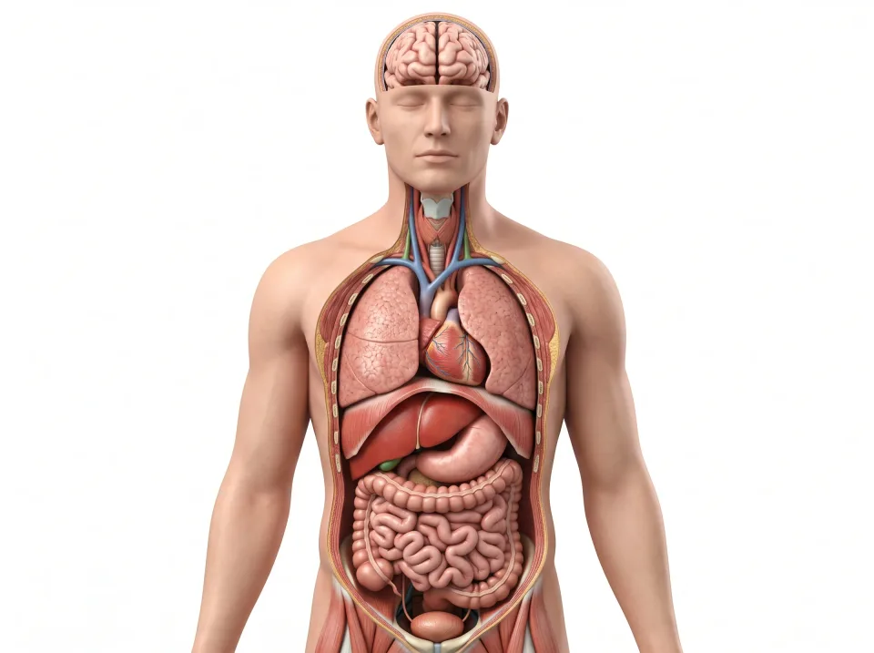
Body location map
Highlighted structure
Heart
Where: Between the lungs in the thorax
Job: Pumps blood through pulmonary and systemic circuits.
Drag to turn. Pinch to zoom. Select a visible structure to learn about it.
Where it sits on a slice
Axial through the heart
Seen from the feet, so the patient's right is on your left.
From cells to a whole person
The body is organized in levels, and each level is built from the one below it. The lowest level is made of molecules, such as water, proteins, fats, sugars, and DNA. Molecules are arranged into , which are the smallest units that are alive. A cell can take in fuel, make energy, build proteins, and respond to its surroundings. The body contains tens of trillions of cells.
A is made up of a group of the same kind of cells, along with the material those cells produce around themselves. In bone, for example, that material is the hard, mineral-filled substance around the bone cells. An is two or more tissues built into a single structure with a shared job. The stomach is a good example. Its lining tissue makes acid, layers of muscle tissue churn, connective tissue holds the organ together, and nervous tissue tells it when to work.
An is a group of organs that cooperate to perform one large task. All the organ systems work together to make a person. There are eleven organ systems: integumentary (the skin), skeletal, muscular, nervous, endocrine, cardiovascular, lymphatic, respiratory, digestive, urinary, and reproductive.
No system works by itself. The lungs load oxygen into the blood, the heart and blood vessels carry it to the tissues so every cell can use it. When one system fails, the others feel the effect within minutes.
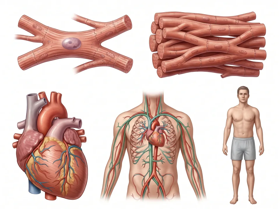
1.Cell
A cardiac muscle cell is one living unit specialized for contraction.
2.Tissue
Many cardiac muscle cells form muscle tissue, with supporting material and other cells.
3.Organ
The heart combines muscle, connective, epithelial, and nervous tissues into a pump.
4.Organ system
The heart and blood vessels cooperate as the cardiovascular system.
5.Person
The cardiovascular system works with all the other systems to sustain one person.
Each view uses a different scale, from microscopic cell to whole person.
Cells, and how the body keeps itself steady
Every cell is wrapped in a thin double layer of fat molecules called a . The membrane is selectively permeable, which means some substances pass freely, some substances are blocked, and some substances can only get through with the help of a protein doorway.
Cells sit in , which fills the narrow spaces between them like grout between tiles. Oxygen and nutrients leave the smallest blood vessels, known as the capillaries, cross this fluid, and enter the cells. At the same time, waste travels in the opposite direction. Most of this exchange happens by , where particles drift from areas of high concentration to areas of low concentration, at no energy cost.
means keeping the body's internal conditions within a workable range. The body controls its temperature like a thermostat set to 37 degrees Celsius, and the value it defends is called the .
When temperature drifts from the set point, the body pushes it back. If you overheat, you sweat, and the vessels in your skin widen so the blood can carry heat out to the surface. If you cool down, you shiver instead. A response that opposes the change is called . It runs almost every control loop in the body, blood sugar and blood pressure included.
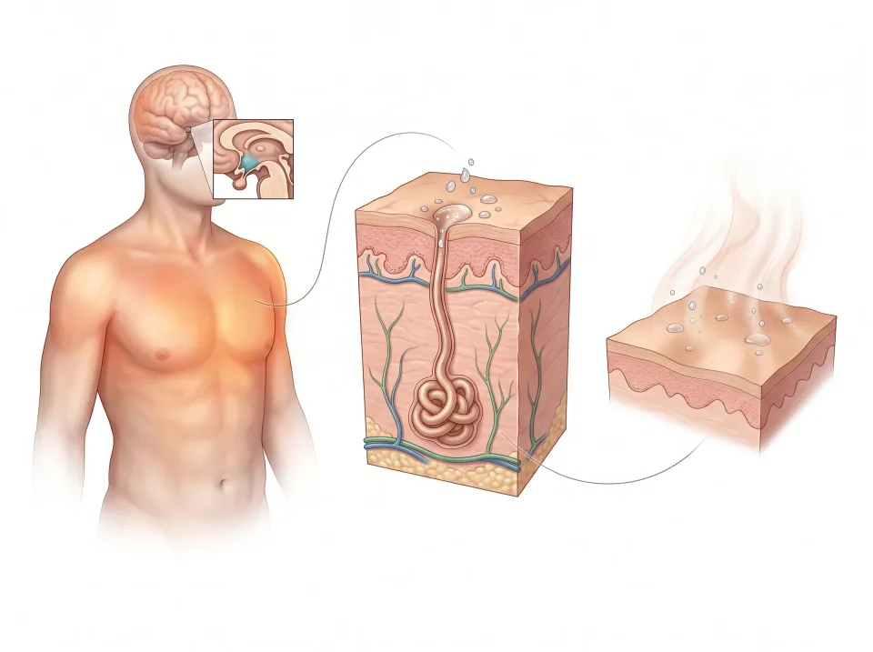
1.Temperature rises
A change is detected: body temperature rises above its usual working range.
2.Sweating increases
Control centers signal sweat glands. Sweat reaches the skin surface.
3.Body cools
Evaporation removes heat. As temperature returns toward its usual range, the response decreases.
The skin views are magnified. Evaporation of sweat removes heat.
Four kinds of tissue
Every organ in the body is built from four tissue types: epithelial, connective, muscle, and nervous.
covers and lines the body's surfaces, inside and out. Its cells are packed tightly into a continuous sheet with little between them. Epithelium forms the surface of your skin, the moist lining inside your cheek, and the inner surface of your stomach and blood vessels. The glands that make sweat and saliva are epithelium too.
supports, binds, cushions, and transports. Its cells are scattered in a large amount of material they produce around themselves, called matrix. The matrix is hard in bone, rubbery in cartilage, soft in fat, and liquid in blood. In a tendon, like the one posterior to your ankle joint, it is tough and stringy.
shortens to produce force, and it comes in three kinds. Skeletal muscle is the kind you control, such as your calf when you stand on your toes. Cardiac muscle is found only in the heart. Smooth muscle works in the walls of the gut, blood vessels, and bladder without conscious effort.
carries electrical signals. Its neurons work like wires, and supporting cells called glia hold and feed them. The tingle that shoots to your little finger when you bump your elbow is nervous tissue at work.
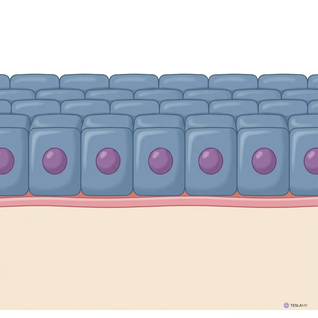
Epithelial tissue
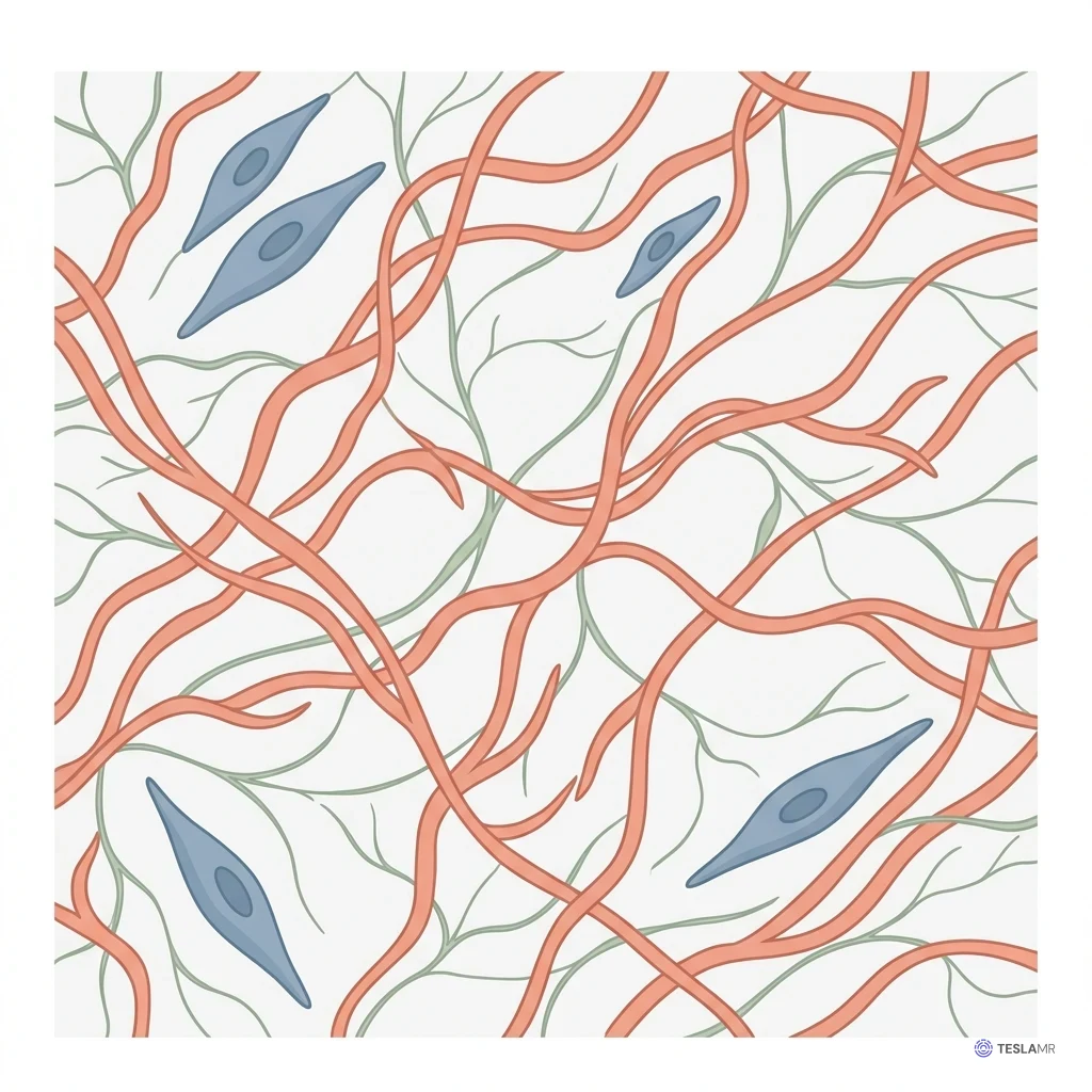
Connective tissue
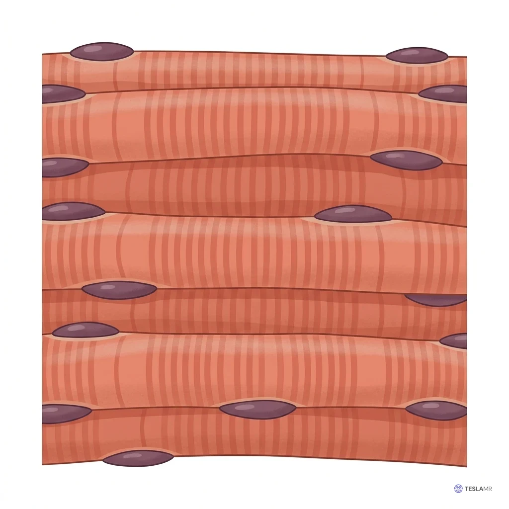
Muscle tissue
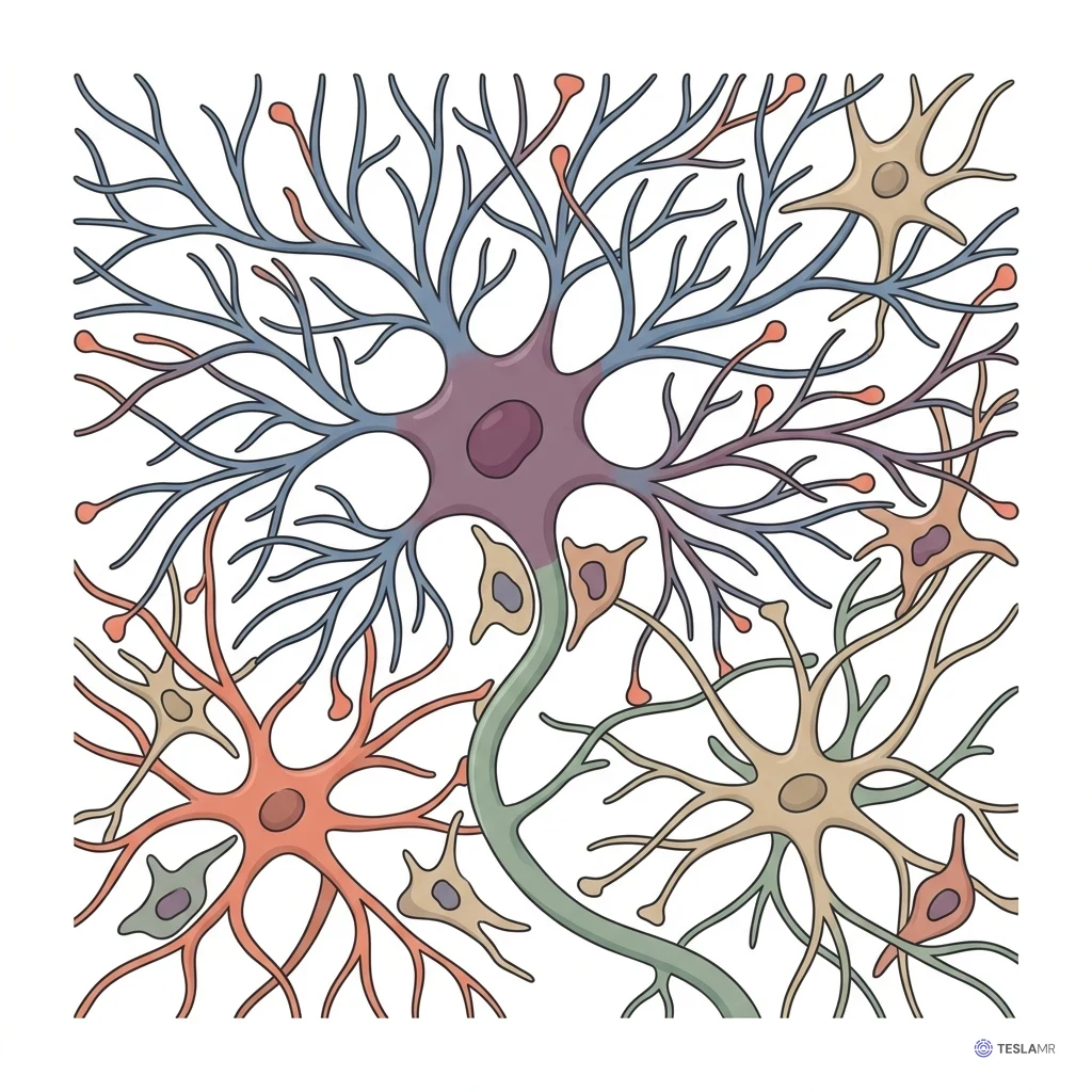
Nervous tissue
Skin, the organ you can see
The is the largest organ in the body and the easiest one to study, because you can see and touch it. Going from superficial to deep, you cross three layers.
The is the most superficial layer, a thin barrier of tightly packed epithelial cells. Its surface cells are dead, flattened, and filled with a tough protein called keratin. The epidermis has no blood vessels of its own, so a shallow scrape turns pink but does not bleed.
The , deep to the epidermis, is connective tissue packed with blood vessels, nerve endings, sweat glands, and hair roots. Because of those vessels and nerves, a cut that bleeds or hurts has reached the dermis.
The lies deep to the dermis and is mostly fat, which cushions the body, insulates it, and stores energy. On many MRI images, this fat shows as a bright rim just deep to the skin, a useful first landmark for telling outside from inside. Some scans are set up to make fat dark instead.
The skin does four jobs at once. As a barrier, it keeps water in and keeps microbes and injury out. It senses touch, pressure, and temperature through the nerve endings in the dermis. It also helps cool the body by sweating and by widening the vessels in the dermis, so heat can escape through the surface.
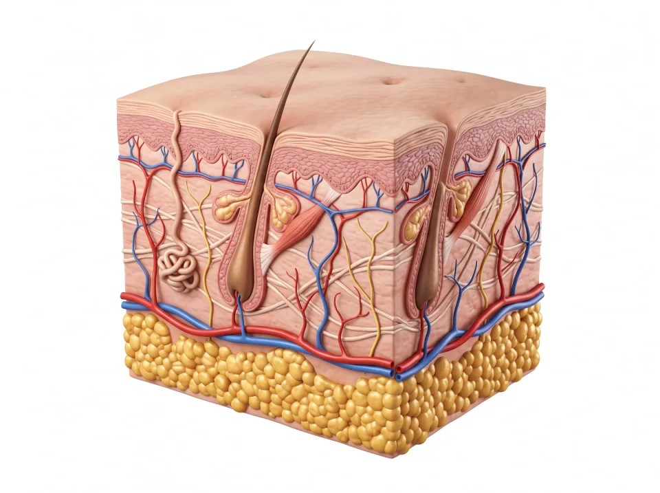
1.Epidermis
Outer barrier. It has no blood vessels.
2.Dermis
Supportive tissue containing blood vessels, nerves, and glands.
3.Subcutaneous layer
Fat below the skin cushions, insulates, and stores energy.
Subcutaneous tissue lies below the skin. The epidermis itself contains no blood vessels.
Cavities and the membranes that line them
Body cavities are enclosed spaces holding the organs of the head and trunk. The in the skull holds the brain. At the base of the skull it opens into the vertebral canal, which runs down the spine and holds the spinal cord.
The is the chest. A lung fills each side, and the between them holds the heart, great vessels, trachea, and esophagus. The floor of the chest is the , a dome of muscle that also drives breathing.
The lies inferior to the diaphragm and holds the stomach, liver, pancreas, kidneys, and most of the intestines. The lies inferior to it, inside the pelvic bones. It holds the bladder, rectum, and reproductive organs: the uterus and ovaries in a female, or the prostate in a male.
There is no wall between the abdominal and pelvic cavities. The boundary is an imaginary line across the top of the pelvic bones, and together they form the .
, thin double-layered sheets, line the thoracic and abdominopelvic cavities. The visceral layer lies on the organ, and the parietal layer lines the cavity wall. Slippery fluid between the layers lets organs slide as you breathe.
The wraps each lung, the wraps the heart, and the lines the abdominopelvic cavity and covers most of its organs. The kidneys and pancreas lie posterior to the peritoneum, against the posterior abdominal wall, and are called .
The meninges wrap the brain and spinal cord instead, protecting and cushioning them.
On a sagittal image near the midline, the cavities stack like the floors of a building: the brain in the cranial cavity at the top, the heart in the chest superior to the dome of the diaphragm, and the abdominal organs inferior to it, continuing without a wall to the bladder posterior to the pubic bone.
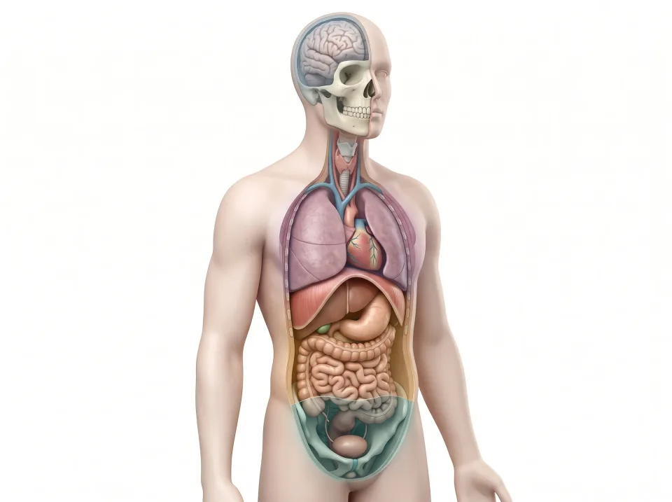
1.Cranial
Inside the skull: the brain.
2.Thoracic
Above the diaphragm: lungs and heart.
3.Abdominal
Below the diaphragm: digestive organs and kidneys near the back.
4.Pelvic
Within the pelvis: bladder, rectum, and internal reproductive organs.
Key ideas to remember
- describes what a structure is and where it sits, and describes what it does.
- Cells build tissues, tissues build organs, organs build systems, and eleven systems build a person.
- Every organ is made from four tissue types: , , , and .
- The brain sits in the cranial cavity, the heart and lungs sit in the thoracic cavity superior to the , and the abdomen and pelvis form one continuous space lined by the .
Check what you learned
Twenty-five questions from this chapter, two of them illustrated, each with the answer, the reasoning, and a memory aid.